Grupo: DANAHER

Excertos do catálogo

THE VERTICAL TURN Leica TCS SP8 DLS Confocal and Digital LightSheet Microscope
Abrir o catálogo na página 1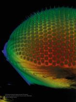
Color coded three-dimensional image of eye specific GFP expression (3P3 promoter) in Drosophila melanogaster. Specimen courtesy of Nadja Dinges, Roignant Lab, IMB Mainz, Germany.
Abrir o catálogo na página 2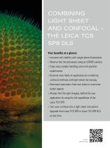
COMBINING LIGHT SHEET AND CONFOCAL – THE LEICA TCS SP8 DLS Your benefits at a glance: > Increase cell viability with single plane illumination > bserve fast live processes using an sCMOS-camera O > njoy easy sample handling and multi-position E experiments > Discover new fields of application by combining confocal methods with light sheet microscopy > Illuminate specimens from two sides to overcome darker regions > lways find the right imaging method for your A application by using the full capabilities of the Leica TCS SP8 > urn your confocal into a light sheet instrument: T Upgrade from...
Abrir o catálogo na página 3
VERTICAL TURN, EXPANDED OPTIONS Our Leica TCS SP8 DLS combines a complete confocal microscope with gentle single plane illumination in one system. The innovative TwinFlect mirror has turned the Leica TCS SP8 confocal platform into an easy and versatile light sheet microscope. Benefit from THE VERTICAL TURN! More options for your research The innovative TwinFlect technology Living specimens – particularly developmental processes in embryos – are often Light sheet microscopy usually requires a very sensitive to light exposure. Illuminating your sample only in one single plane at dedicated...
Abrir o catálogo na página 4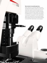
High-end optics for demanding applications Superior objectives for a wide range of applications is one of Leica Microsystems’ hallmarks. The heart of our system turning the light sheet vertically is formed by the objectives and the TwinFlect mirror. With the choice of two different illumination objectives, the Leica HC PL Fluotar 2.5x/0.07 or the Leica HCX PL FLUOTAR 5x/0.15, you can shape the light sheet depending on the experiment's requirements. In order to reveal the finest details or have a larger field of view, you can pick the optimal detection objective, either the Leica HC FLUOTAR...
Abrir o catálogo na página 5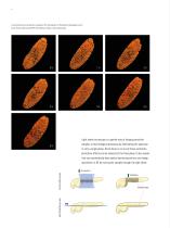
Low phototoxicity and specimen imaging in 3D: Development of Drosophila melanogaster over 6 hours. Probe: Light-sensitive RFP. 3D rendering. 150 µm z stack, 30 sec/stack. Light sheet microscopy is a gentle way of imaging sensitive samples or fast biological processes by illuminating the specimen in only a single plane. Since there is no out-of-focus excitation, phototoxic effects can be reduced to the focal plane. It also means that you automatically have optical sectioning and you can image specimens in 3D by moving the sample through the light sheet. Light Sheet Microscopy Confocal...
Abrir o catálogo na página 6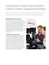
CONDUCT AND DOCUMENT LONG-TERM OBSERVATIONS Light sheet technology by Leica Microsystems allows you to observe the development of interesting organisms over a longer span of time using low illumination and high speed. Watching developments in real time and 3D Imaging requires light, but too much light can damage your cells. Light sheet microscopy is the most gentle imaging method to date, as it reduces the overall photodamage from phototoxicity and bleaching. This automatically increases the viability of your specimen. Particularly researchers in developmental biology benefit from light...
Abrir o catálogo na página 7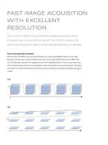
FAST IMAGE ACQUISITION WITH EXCELLENT RESOLUTION Do you want to observe fast and periodic biological processes in three dimensions over an extended time period? Then the DLS module is the right choice. Discover the beauty of a beating zebrafish heart, for example. Track very fast periodic movements With the Leica TCS SP8 DLS you can choose between two cutting-edge sCMOS cameras for fast image acquisition with excellent resolution and high frame rates: The pco.edge sCMOS camera and the ORCA-flash 4.0 V2 by Hamamatsu are both fully integrated into the LAS X LightSheet Wizard. A new xytz scan...
Abrir o catálogo na página 8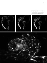
High-speed imaging: Beating zebrafish heart, transgenic line flik1::EGFP. Top: transverse section at different time points. Bottom: Post-acquisition alignment after xytz recording. Recording speed: 120 frames/second. Courtesy of Emily Steed, Vermot Lab, IGBMC Strasbourg, France.
Abrir o catálogo na página 9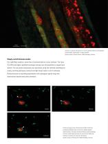
3:23:00.000 Long-term, multicolor observations: 17 hours of zebrafish lateral line development. Zebrafish 36hpf, CldnB:lynGFP / Cxcr4B:nuclearRFP. Sample courtesy of Darren Gilmour, EMBL Heidelberg, Germany. Simply switch between modes Our LightSheet module is more than a functional add-on to your confocal. The Leica TCS SP8 and Digital LightSheet synergize and give you the possibility to expand your options. You can easily manipulate your specimens using the confocal technology by simply switching between confocal and light sheet mode in LAS X software. Photoconversion or wounding...
Abrir o catálogo na página 10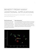
BENEFIT FROM MANY ADDITIONAL APPLICATIONS We have added light sheet technology to our confocal platform – you get two high performance microscopes in one. Always the right laser option Familiar sample preparation All visible lasers of your Leica TCS SP8 Due to the vertical experimental setup of the Leica TCS SP8 DLS, you can stick closely confocal are ready to be used for light to your routine sample preparation. The specimens are mounted in conventional glass sheet imaging. The highest flexibility in bottom petri dishes and are directly accessible. You even have the possibility of choosing...
Abrir o catálogo na página 11
PARTNERS IN SCIENCE – WE FOCUS ON YOUR NEEDS Time is too precious to spend on complicated experimental set ups. That is why we focus on intuitive and convenient system design. Workflow-oriented software design The LAS X microscope software guides users step by step through data recording and evaluation. The workflow-oriented design helps you to use the instrument more efficiently. A convenient calibration routine establishes the light sheet precisely. Dual-sided illumination of the sample comes by design: Each of the two opposing mirrors of the TwinFlect can be targeted by the scanner to...
Abrir o catálogo na página 12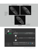
Side opposite of illumination suffers from blurring Fused image with superior quality LightSheet Filter: Detection Objective:
Abrir o catálogo na página 13Todos os catálogos e folhetos técnicos Leica Microsystems GmbH
-
 Mateo FL Flyer EN
2 Páginas
-
 UC Enuity Product-Brochure EN
8 Páginas
-
 DVM6 Brochure en
16 Páginas
-
 Leica M50, M60, M80
12 Páginas
-
 XL Stand
4 Páginas
-
 LED1000
16 Páginas
-
 LED3000 BLI
20 Páginas
-
 LED5000 NVI
20 Páginas
-
 LED3000 NVI
20 Páginas
-
 LED3000 DI
20 Páginas
-
 LED5000 HDI
20 Páginas
-
 LED5000 CXI
20 Páginas
-
 LED2500
8 Páginas
-
 LED5000 MCI
20 Páginas
-
 LED3000 MCI
20 Páginas
-
 LED5000 SLI
20 Páginas
-
 LED3000 SLI
20 Páginas
-
 LED2000
8 Páginas
-
 LED5000 RL
20 Páginas
-
 LED3000 RL
20 Páginas
-
 Water Immersion Micro Dispenser
2 Páginas
-
 EL6000
4 Páginas
-
 M320 F12 for ENT
12 Páginas
-
 M620 TTS
4 Páginas
-
 M220 F12
8 Páginas
-
 RUV800
4 Páginas
-
 DI C800
2 Páginas
-
 M620 F20
8 Páginas
-
 M822 F40 / F20
20 Páginas
-
 M844 F40 / F20
16 Páginas
-
 Proveo 8
16 Páginas
-
 Envisu
8 Páginas
-
 EnFocus
8 Páginas
-
 M525 F20
12 Páginas
-
 FL400
FL4004 Páginas
-
 Leica M525 OH4
20 Páginas
-
 Leica M720 OH5
24 Páginas
-
 Leica M530 OH6
16 Páginas
-
 Leica M530 OHX
16 Páginas
-
 Leica CaptiView
4 Páginas
-
 Leica GLOW800
4 Páginas
-
 Leica ARveo
16 Páginas
-
 Leica PROvido
8 Páginas
-
 LAS X Steel Expert
4 Páginas
-
 Leica LMD Software
16 Páginas
-
 Leica Map
12 Páginas
-
 Leica Cleanliness Expert
4 Páginas
-
 Leica LAS X Industry
4 Páginas
-
 Leica LAS X Life Science
4 Páginas
-
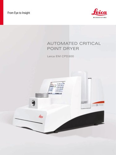 Leica EM CPD300
12 Páginas
-
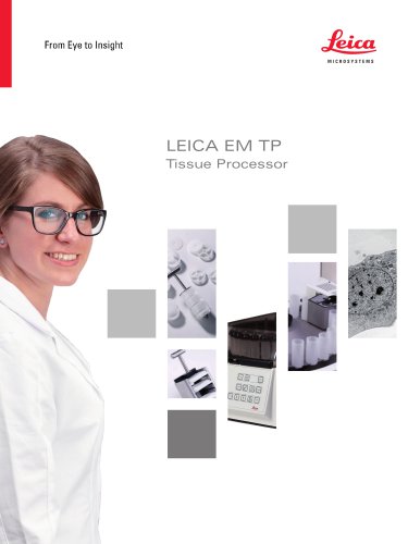 Leica EM TP
4 Páginas
-
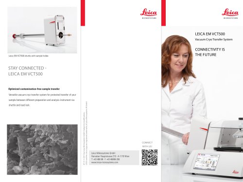 Leica EM VCT500
2 Páginas
-
 Leica EM ACE900
12 Páginas
-
 Leica EM ACE200
12 Páginas
-
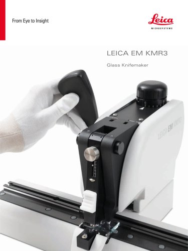 Leica EM KMR3
8 Páginas
-
 Leica EM AFS2
8 Páginas
-
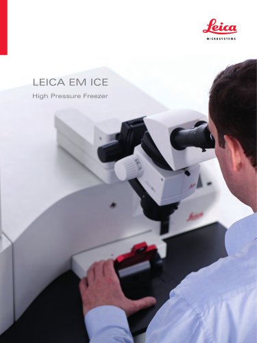 Leica EM ICE
12 Páginas
-
 Leica LIGHTNING
2 Páginas
-
 Leica SP8 DIVE
20 Páginas
-
 Leica K5
2 Páginas
-
 Leica DFC295
6 Páginas
-
 Leica DFC3000 G
6 Páginas
-
 Leica EC4
4 Páginas
-
 Leica DFC7000 T / DFC7000 GT
4 Páginas
-
 Leica DFC9000
2 Páginas
-
 Leica MZ10 F
4 Páginas
-
 Leica M165 FC
16 Páginas
-
 THUNDER Imager Model Organism
2 Páginas
-
 Leica DM IL LED
12 Páginas
-
 Leica DMi1
2 Páginas
-
 THUNDER Imager Tissue
2 Páginas
-
 Leica FS M
12 Páginas
-
 Leica FS C
20 Páginas
-
 Leica FS CB
20 Páginas
-
 Leica DM3000 / DM3000 LED
16 Páginas
-
 Leica DM750
12 Páginas
-
 Leica DM500
12 Páginas
-
 Leica DM300
8 Páginas
-
 Leica DM1750 M
12 Páginas
-
 Leica FS4000 LED
20 Páginas
-
 Leica DM2000 / DM2000 LED
16 Páginas
-
 Leica DM1000
16 Páginas
-
 Leica DM1000 LED
16 Páginas
-
 Leica DM2500 & DM2500 LED
16 Páginas
-
 Leica DM4 B & DM6 B
16 Páginas
-
 Leica DMC2900
6 Páginas
-
 Leica DMC6200
8 Páginas
-
 Leica DMC5400
4 Páginas
-
 Leica Z6 APO
16 Páginas
-
 Leica Z16 APO
16 Páginas
-
 Leica M205 FCA / M205 FA
16 Páginas
-
 Leica DM12000 M
8 Páginas
-
 Leica DM8000 M
8 Páginas
-
 Leica DM4 P, DM2700 P, DM750 P
12 Páginas
-
 Leica EM ACE600
12 Páginas
-
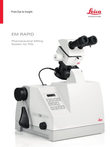 Leica EM RAPID
8 Páginas
-
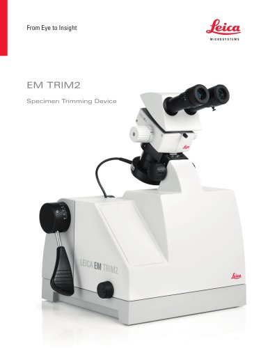 Leica EM TRIM2
8 Páginas
-
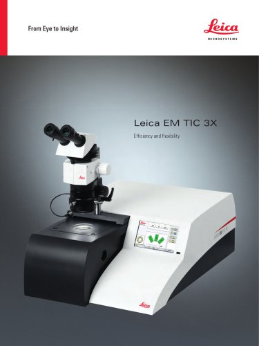 Leica EM TIC 3X
16 Páginas
-
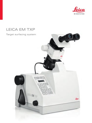 Leica EM TXP
10 Páginas
-
 Leica DM2700 M
12 Páginas
-
 DMi8 M / C / A
12 Páginas
-
 Leica A60 S / Leica A60 F
16 Páginas
-
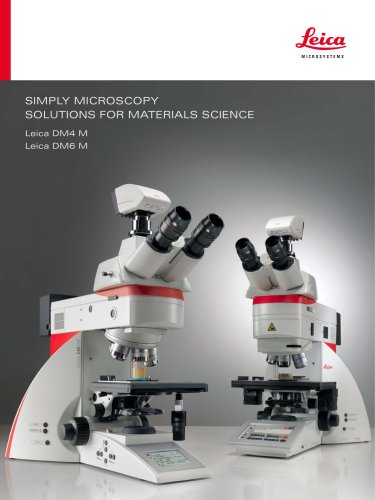 SOLUTIONS FOR MATERIALS SCIENCE
12 Páginas
-
 Leica StereoZoom line
20 Páginas
-
 TL-Bases
12 Páginas
-
 TCS SP8
20 Páginas
-
 TCS SP8 STED 3X
24 Páginas
-
 TCS SP8 Objective
24 Páginas
-
 AOBS
AOBS16 Páginas
-
 Leica DM3 XL
7 Páginas
-
 Leica DMi8 S
12 Páginas
-
 Leica M50/M60/M80
12 Páginas
-
 Leica DM750 M
12 Páginas
-
 Leica DM4 M/ DM6 M
12 Páginas
-
 Leica A60 S/A60 F
16 Páginas


