Catalog excerpts

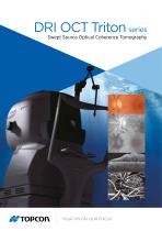
DRI OCT Triton series Swept Source Optical Coherence Tomography
Open the catalog to page 1
See. Discover. Explore. The diagnostic power of Swept Source OCT Deep Range Imaging. Swept Source adds a new dimension to OCT. The Topcon DRI OCT Swept Source is easy to use, provides unique clinical information, and has improved my practice. For the first time, we can in-vivo visualize not only the vitreo-retinal interface but also the cortical vitreous which is important at the time when more and more therapies are delivered via intra-vitreal injections. Deeper imaging brings choroidal thickness, helping guide my clinical decisions. Seeing more helps guide my therapy and allows me to...
Open the catalog to page 2
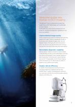
Welcome to the new frontier in OCT imaging The DRI OCT Triton combines the world's first Swept Source OCT technology with multimodal fundus imaging. Multimodal All-in-One fundus imaging tool will bring the next level of diagnostic capability to you and your patients. Unprecedented image quality Triton's Swept Source with its extremely fast scanning speed and longer 1,050nm wavelength results in stunningly clear, detailed images, even into the deepest layers of the eye with short acquisition time. You will not only see the retina and vitreous, but also the choroid and the sclera like never...
Open the catalog to page 3
See deeper. See more. Proliferative diabetic retinopathy Lateral: 12mm Courtesy: rof. P. E. Stanga, Manchester Royal Eye Hospital, Manchester Vision Regeneration (MVR) Lab at P NIHR / Welcome Trust Manchester CRF & University of Manchester Courtesy: rof. P. E. Stanga, Manchester Royal Eye Hospital, Manchester Vision Regeneration (MVR) Lab at P NIHR / Welcome Trust Manchester CRF & University of Manchester * FA photography and FAF photography can be performed using only DRI OCT Triton plus
Open the catalog to page 4
Central serous retinopathy Pro--macula1 nn'.torior tfetdfhmenr Rur^.i prem1cul1rli Swoflen photoreceptor outer segments Slip I olio'll IhmS rxEcM^'iing from tO1CO to Optic rsOftre I'li'.'nJ Merw surf nee of sclera Courtesy: Prof. P. E. Stanga, Manchester Royal Eye Hospital, Manchester Vision Regeneration (MVR) Lab at NIHR / Welcome Trust Manchester CRF & University of Manchester Courtesy: Prof. P. E. Stanga, Manchester Royal Eye Hospital, Manchester Vision Regeneration (MVR) Lab at NIHR / Welcome Trust Manchester CRF & University of Manchester FA photography and FAF photography can be...
Open the catalog to page 5
Pathological myopia Sbbretinad choroidal neovascular merr-brane ViTfGomaculaf atbaclrme-ni Fovtas Cystic changes Thin Choroid Reiro-oculai op-seiefa? highly reflective tissue Lateral: 12mm Courtesy: Prof. P E. Stanga, Manchester Royal Eye Hospital, Manchester Vision Regeneration (MVR) Lab at NIHR / Welcome Trust Manchester CRF & University of Manchester Macular pucker Retinal distort ion itfltf thickening Epiretinaf nraimlsrwii iiKiratiriAi cyatold chan Di-vtortiofl of Ellipsoid layer Lateral: 12mm Courtesy: Prof. P. E. Stanga, Manchester Royal Eye Hospital, Manchester Vision...
Open the catalog to page 6
Image through cataract a), b), c) Courtesy: Kazuya Yamagishi, MD (Hirakata Yamagishi Eye Clinic, Japan)
Open the catalog to page 7
Swept Source takes OCT technology to a whole new dimension. Envision the possibilities DRI OCT Triton’s Swept Source OCT technology and reduces risks of light attenuation by cataract and long wavelength 1,050nm light enable both a deeper vitreous opacity, making OCT imaging more feasible imaging range and a better tissue penetration, for the patients with those diseases. Advantages of compared with the conventional spectral domain DRI OCT Triton’s technology improvement over the OCT. The OCT images captured by DRI OCT Triton conventional spectral domain OCT will provide more are clearly...
Open the catalog to page 8
Swept Source OCT Angiography | y utilizing cutting-edge Swept Source technology B with a 1,050nm wavelength, high-quality OCT imaging technique to visualize the microvascular Angiography images are acquired | asier recognition of abnormalities by using layer E network. It is now available any time you need it. The optional OCT Angiography module offers by layer “tissue peeling” intuitive graphical user non-invasive observation of the microvascular interface | mproved patient comfort*4 - no dyes or dilation I structures reducing the need for conventional fluorescein angiography. required,...
Open the catalog to page 9
Improved clinical efficacy with sophisticated analysis functions. En Face image Projection image En Face imaging allows for independent dissection of the vitreoretinal interface, retina, retinal pigment epithelium (RPE), and choroid by flattening B-scan image. Pathology throughout the posterior pole can be studied and correlated with a patient’s symptoms, their abnormality, and its progression. Lamina Cribosa Lamina Cribosa Courtesy: Prof. T. Nakazawa, Tohoku University, Japan Original Image Flattened image Courtesy: Prof. T. Nakazawa, Tohoku University, Japan To visualize vitreous Dynamic...
Open the catalog to page 10
Normative database with Swept Source OCT DRI OCT Triton includes a normative database for statistical comparison of the thickness maps and parameters. By comparing individual measurement value with the corresponding normative range, the DRI OCT Triton provides you with a powerful reference tool to enhance your analysis in both research and patient diagnosis. 7 boundaries segmentation/5 layers thickness map/caliper function Retinal tissue layers are automatically segmented by the Topcon Advanced Boundary Software (TABSTM), enabling to quantify the internal thickness for change analysis....
Open the catalog to page 11
DRI meets multimodal fundus imaging: see the whole picture. Swept Source OCT incorporates multimodal fundus imaging DRI OCT Triton can acquire the OCT and fundus image in a single capture. Pinpoint RegistrationTM identifies the location of B-scan on the fundus image. Clear comparison between the B-scan and fundus image can support clinical efficiency during diagnosis. OCT + Color fundus High quality fundus images The DRI OCT Triton offers a color, non-mydriatic fundus image. Fundus Angiography (FA) and Fundus Autofluorescence (FAF) are available to meet your needs. The all in one device...
Open the catalog to page 12All TOPCON EUROPE POSITIONING catalogs and technical brochures
-
RL-SV2S
2 Pages
-
FC-6000
4 Pages
-
3D CONSTRUCTION
32 Pages
-
2D / 3D Excavator Control
2 Pages
-
IS-1 Series
9 Pages
-
FS-1 series
4 Pages
-
IMAGEnet i-base
2 Pages
-
OMS-800 Series
8 Pages
-
SP-1P
8 Pages
-
KR-1W
2 Pages
-
CA-800
16 Pages
-
SL-D Series
16 Pages
-
SL-2G
4 Pages
-
SL-D701
2 Pages
-
SL-D701
8 Pages
-
TRC-50DX
8 Pages
-
TRC-NW8 series
8 Pages
-
3D OCT
12 Pages
-
GT SERIES
4 Pages
-
GR-5
4 Pages
-
3D-MC MAX
4 Pages
-
X-53
2 Pages
-
X-53i
2 Pages
-
DynaRoad
4 Pages
-
Topcon Tierra
4 Pages
-
DL series
4 Pages
-
Robotic Total Stations
4 Pages
-
Dozer GPS + Control
4 Pages
-
Dozer LPS control
4 Pages
-
MACHINE CONTROL CATALOGUE
16 Pages
-
Dozer Laser Control
4 Pages
-
LASER CATALOGUE
12 Pages
-
3D Mobile Mapping System
6 Pages
-
ScanMaster - Data Management
4 Pages
-
TopSURV7
4 Pages
-
Topcon Tesla RTK
4 Pages
-
IP-S2 HD
4 Pages
-
IP-S2 Compact
8 Pages
-
ImageMaster
4 Pages
-
Survey / Mapping CATALOGUE
28 Pages
-
IS Imaging Station
4 Pages
-
Field Controller (FC-250)
4 Pages
-
Laser Scanner (GLS-1500)
4 Pages
Archived catalogs
-
Crossline Laser (LC-2,LC-4X)
4 Pages
-
Laser Range
8 Pages
-
GPS+Receivers (HiPer Pro)
4 Pages
-
GPS+Receivers (Hiper Series)
6 Pages
-
Leaflet FC-200
2 Pages
-
Leaflet ATG-Series
4 Pages


