
Catalog excerpts

3D OCT-1 Maestro Optical Coherence Tomography
Open the catalog to page 1
Discover the OCT world at your fingertips
Open the catalog to page 2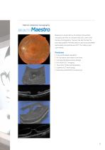
Optical coherence tomography Based on a long history of product innovation, including the first to combine SD OCT with color fundus photography, Topcon has set the bar for providing patient friendly, easy to use and completely automated comprehensive OCT for today's eye care needs. Features | Fully automated operation | Rich analysis and report functions | Compact & space saving design | EN VIEW OCT Imaging | True color fundus photography | Superb OCT technology | Network and DICOM connectivity
Open the catalog to page 3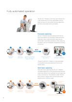
Fully automated operation The 3D OCT- Maestro is the most user-friendly OCT in the market due to its fully automated function. With one touch on the screen, the auto alignment, auto focus and auto shoot is activated. Full-Auto capturing 3D OCT-1 Maestro requires nothing more than to touch the capture icon and [Start Capture] button. Alignment, focus, optimizing and capturing are per or ed in automatic procedure. After capturing, f m the report can be immediately displayed by clicking on the icon. 1 Registering patient Selecting a capture icon Adjusting the chinrest position and touching...
Open the catalog to page 4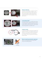
Manual capturing Depending on pathology or on patient’s condition, automatic scanning shall be avoided. In such cases, manual mode will help to adjust alignment and scan ing position. Variety of functions are n available and all are smoothly operated on touch panel monitor. Live Fundus View™ Live Fundus View (OCT-LFV) is a perfect tool for cap u t ring small pupils with a diameter of ø2,5mm. OCT-LFV is a live projection image with reflection at the retina. It gives a clear live fundus image. Disc, retinal vessels and scanning position is very easy to see. Cataract mode In case there is...
Open the catalog to page 5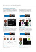
Rich analysis and report functions Rich analysis and report functions The 3D OCT-1 Maestro allows for rich analysis functions Reports contain for example optic disc analysis, for Macula, Glaucoma or Anterior. Comprehensive, 3D macula analysis, 12 mm 3D wide scan and predefined report templates allow you to see and others. The anterior analysis is an option on the print diagnostic output in a clear way. 5 Line cross scan This scans with 5 line scan horizontally and vertically Horizontal box scan in macula area. 3D imaging is in an instant. This is useful for screening and for useful to...
Open the catalog to page 6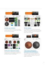
This allows to screen from the fovea to the optic Disc topography which combines fundus photo nerve by single scanning. Thickness maps of RNFL, graphy, various peripapillary parameters and RNFL thickness is available. The normative database for RNFL is also incorporated. Vertical box scan in macula area. GCC analysis is Color fundus photography/ peripheral fundus photography available and normative database for RNFL, GCC Non mydriatic color fundus photography is and retina thickness is incorporated. possible. The report template is ready for color 3D Macula (V) glaucoma analysis fundus...
Open the catalog to page 7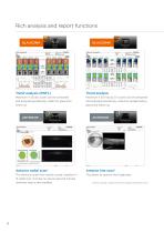
Rich analysis and report functions GLAUCOMA Trend analysis (RNFL) Trend analysis Maximum 4 3D disc scans can be compared Maximum 4 3D macula (V) scans can be compared and analyzed periodically. Useful for glaucoma and analyzed periodically. Useful for preperimetory glaucoma follow-up. Anterior radial scan* Anterior line scan* This allows to check the central cornea condition in This allows to observe the Angle area. 12 radial scan. Corneal curvature map and corneal thickness map is also available. *Anterior scanning is optional with anterior segment attachment (HA-2).
Open the catalog to page 8
Compact & space saving design Compact & space saving design Due to the rotatable touch screen control panel, the operator can use the 3D OCT-1 Maestro from several positions: the classic position, positioned behind the patient and from the side. This results in a superb patient interaction and a space saving set up. The compact design and small footprint of the 3D OCT-1 Maestro allows it to be installed on a refraction unit or a table like Topcon's IC-1. Front position Brightness shield BS-1 The optional Topcon BS-1 Brightness Shield will help to reduce undesired light. The slim...
Open the catalog to page 9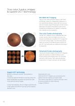
EN VIEW OCT Imaging Topcon's EN VIEW software, based on EN FACE technology, allows for independent dissection of the vitreoretinal interface, retina, REP and choroid and will uniquely project these layers so that macula pathology throughout the posterior pole can be studied and correlated with a patient's symptoms, their disease and its progression. True color fundus photography The 3D OCT-1 Maestro has an integrated full color fundus camera. With one finger touch you can acquire simultaneously a posterior OCT image and a true color fundus image. This real fundus photo helps you quickly to...
Open the catalog to page 10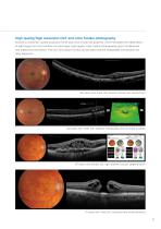
High quality/high resolution OCT and color fundus photography 50,000 A-scans/sec. speed produces fine B-scan and smooth 3D graphics, which facilitates the observation of pathology form and condition on each layer. High quality color fundus photography gives fundamental and additional information. The OCT and colour fundus can be said to be the inseparable combination for daily diagnosis. 85-years old, male, OD, branch retinal vein obstruction 62-years old, male, OS, diabetic retinopathy and circinate exudate 97-years old, female OD, age related macular degeneration 71-years old, male OD,...
Open the catalog to page 11
Specifications Observation & photography of fundus image*** Scan mode Color, red-free* Picture angle 45°/30° or equivalent (digital zoom) 34,8mm (in fundus photography) 62,6mm (in anterior segment photography**) Operating distance Photographable Diameter of pupil 45°: Ø 4.0mm or more Small pupil diameter: Ø 3.3mm or more Observation & photographing of the fundus/ anterior segment tomogram (on fundus) Horizontal direction 3 ~ 12mm, Vertical direction 3 ~ 9mm Scan range Horizontal direction 3 ~ 6mm, Vertical direction 3 ~ 6mm Scan speed Lateral resolution In-depth resolution Photographable...
Open the catalog to page 12All TOPCON EUROPE POSITIONING catalogs and technical brochures
-
 RL-SV2S
2 Pages
-
 FC-6000
4 Pages
-
 3D CONSTRUCTION
32 Pages
-
 2D / 3D Excavator Control
2 Pages
-
 IS-1 Series
9 Pages
-
 FS-1 series
4 Pages
-
 IMAGEnet i-base
2 Pages
-
 OMS-800 Series
8 Pages
-
 SP-1P
SP-1P8 Pages
-
 KR-1W
KR-1W2 Pages
-
 CA-800
16 Pages
-
 SL-D Series
16 Pages
-
 SL-2G
SL-2G4 Pages
-
 SL-D701
2 Pages
-
 SL-D701
8 Pages
-
 TRC-50DX
8 Pages
-
 TRC-NW8 series
8 Pages
-
 DRI OCT Triton series
20 Pages
-
 GT SERIES
4 Pages
-
 GR-5
GR-54 Pages
-
 3D-MC MAX
4 Pages
-
 X-53
X-532 Pages
-
 X-53i
X-53i2 Pages
-
 DynaRoad
4 Pages
-
 Topcon Tierra
4 Pages
-
 DL series
4 Pages
-
 Robotic Total Stations
4 Pages
-
 Dozer GPS + Control
4 Pages
-
 Dozer LPS control
4 Pages
-
 MACHINE CONTROL CATALOGUE
16 Pages
-
 Dozer Laser Control
4 Pages
-
 LASER CATALOGUE
12 Pages
-
 3D Mobile Mapping System
6 Pages
-
 ScanMaster - Data Management
4 Pages
-
 TopSURV7
4 Pages
-
 Topcon Tesla RTK
4 Pages
-
 IP-S2 HD
4 Pages
-
 IP-S2 Compact
8 Pages
-
 ImageMaster
4 Pages
-
 Survey / Mapping CATALOGUE
28 Pages
-
 IS Imaging Station
4 Pages
-
 Field Controller (FC-250)
4 Pages
-
 Laser Scanner (GLS-1500)
4 Pages


Archived catalogs
-
 Crossline Laser (LC-2,LC-4X)
4 Pages
-
 Laser Range
8 Pages
-
 GPS+Receivers (HiPer Pro)
4 Pages
-
 GPS+Receivers (Hiper Series)
6 Pages
-
 Leaflet FC-200
2 Pages
-
 Leaflet ATG-Series
4 Pages


