
- Catalogs
- Shimadzu France
- iMScope Trio


iMScope Trio

iMScope Trio
The iMScope TRIO represents a significant advancement in imaging mass spectrometry by integrating an optical microscope with a mass spectrometer. This combination allows for high-resolution morphological imaging and the identification and visualization of specific molecular distributions, enhancing research capabilities across various fields.
- Multiplexed Imaging: Generates molecular distribution images based on mass spectrometry signal intensity for specific ions.
- Mass Spectrometer: Provides mass spectra at multiple points, allowing for detailed molecular analysis.
- Optical Microscope: Captures high-resolution images, which can be superimposed with mass spectrometry data for comprehensive analysis.
- Medical Research: Useful for biomarker discovery, lipid and metabolite analysis, and pathological studies by identifying disease-related molecules and visualizing their distribution.
- Pharmaceuticals: Assists in pharmacokinetic analysis, drug localization, and toxicity mechanism analysis, providing insights into drug efficiency and safety.
- Food Industry: Evaluates quality and ingredient research of agricultural products, aiding in the inspection of contaminants.
- Cosmetics and Industry: Supports surface analysis and homogeneity evaluation, crucial for product development and quality control.
- High Spatial Resolution: Enables detailed imaging of biological tissues and organs, crucial for pharmacokinetic and toxicity studies.
- Overlaying Images: Combines optical and mass spectrometry images to provide a comprehensive view of molecular localization and morphological changes.
- Experiment Conditions: Detailed parameters such as sample type, matrix used, measurement pitch, and laser diameter are specified for various experiments, ensuring reproducibility and accuracy.
- Cancer Research: Identification of localized molecules in cancerous mouse liver cells, highlighting increased metabolites in metastatic cancer areas.
- Drug Discovery: Visualization of drug distribution in pharmacokinetic studies, demonstrating the retention and localization of drugs like famotidine and paclitaxel in specific tissues.
The iMScope TRIO is a powerful tool for advancing research in multiple disciplines by providing detailed molecular and morphological insights. Its ability to integrate optical and mass spectrometry data makes it invaluable for cutting-edge research and development.
The document discusses the use of imaging mass spectrometry (IMS) in analyzing metabolite distribution in plants and its applications in food and drug development. It highlights the potential of IMS in improving food quality and developing new plant varieties.
IMS is used to visualize metabolite distribution in tomato seedlings and rice endosperm. This technology aids in developing improved plant varieties and analyzing functional metabolites for processed foods and drugs.
IMS is employed in industrial fields for surface inspection, analyzing contaminants on printed circuit boards, and ensuring high-quality production. It provides insights that conventional methods cannot detect.
The iMScope TRIO is a high-performance analysis system combining optical and mass spectrometric images. It offers a 5 μm laser diameter for high-resolution imaging and a high-precision tandem mass spectrometer for structural analysis.
The workflow involves placing a sample, capturing an optical image, applying a matrix, acquiring mass spectra, and visualizing molecular distribution. The system integrates optical and MS images for comprehensive analysis.
The Imaging MS Solution software facilitates efficient statistical analysis, including Hierarchical Cluster Analysis (HCA), Region of Interest (ROI) Analysis, and Principal Component Analysis (PCA). These tools help in identifying and comparing substances in different regions.
The iMLayer system ensures reliable matrix vapor deposition, enhancing ionization consistency. It supports high-resolution imaging and increased signal intensity.
The document concludes with the potential of IMS in biomarker search and distribution analysis, emphasizing its role in advanced research and development.
Catalog excerpts

Imaging Mass Microscope iMScope TRIO
Open the catalog to page 1
Imaging Mass Spectrometry Qualitative Analysis Optical Microscope Introducing the New Era of Imaging Mass Spectrometry Imaging mass spectrometry is a revolutionary new technology. The instrument is a combination of an optical microscope which allows the observation of high-resolution morphological images, with a mass spectrometer which identifies and visualizes the distribution of specific molecules. Superimposing the two images obtained based on these very different principles, has created a significant new research tool, the imaging mass microscope. The accurate and high resolution mass images...
Open the catalog to page 3
Optical microscope Capture an optical microscope image Mass Spectrometer Mass spectra at multiple points Apply matrix to tissue section and irradiate with laser beam. Mass Spectrometry ■■■■■■■■■■■■■ =■■■■■ Multiplexed imaging Generate molecular distribution images (multiplexed imaging) based on ms signal intensity
Open the catalog to page 4
Imaging Mass Spectrometry identifies what you see at the molecular level iMScope TRIO transforms your data from just "Observation" to "Analysis" Optical microscopes cannot determine which molecules are localized in the region of interest. On the other hand, the positional information of molecules is lost in mass spectrometric analysis, where sample extraction and homogenization is needed for sample pre-treatment. What if the qualitative analysis and localization of specific molecules and compositional and morphological observation could be obtained from a single analysis through microscope observation?...
Open the catalog to page 5
Medical Research In medical research, the iMScope TRIO is particularly useful for identifying disease-related molecules (biomarker discovery) and rendering two-dimensional distributions of those substances (visualization). Also biological mechanism analysis and pathological research through identifying the location of target molecules (profiling) can be achieved.
Open the catalog to page 6
Identification of Localized Molecules in Cancerous Mouse Liver Cells The iMScope TRIO makes it possible to obtain information regarding which molecules are localized within the region of interest in an organ, e.g. cancerous area. Until now, biomarker discovery has been carried out by LCMS or other mass spectrometry techniques for each organ. Bright Field Optical Image Fluorescence Optical Image Experiment Conditions Sample: mouse liver Matrix: 9-AA (aminoacridine, sprayed) Measurement pitch: 10 μm Laser diameter: 10 μm Measurement points: 200 × 200 (40,000 points) GSSG 4.0 Contents (μmol/g tissue)...
Open the catalog to page 7
Profiling of Biological Tissue and Organ Cross Sections The iMScope TRIO, which incorporates database searching with MS and MSn data, can be applied not only to visualize the localization of a certain molecule, but also to identify the molecules relating to the cause of disease. Seminolipids in mouse testis Optical Image Experiment Conditions Sample: mouse testis Matrix: 9-AA (aminoacridine, vapor deposited) Measurement pitch: 10 μm Laser diameter: 10 μm Measurement points: 250 × 250 (62,500 points) Measurement time: about 3 hours The images show characteristic distributions of seminolipids (m/z...
Open the catalog to page 8
Visualization of Lipid in a Mouse Cerebellum A wide variety of molecules relate to diseases states. Imaging mass spectrometry using the iMScope TRIO can detect a wide range of molecules within a defined mass range. Therefore, distribution information for several target molecules with different molecular weights can be determined simultaneously during a single measurement. Optical Image Experiment Conditions Sample: mouse cerebellum Matrix: DHB (vapor deposited) Measurement pitch: 10 μm Laser diameter: 10 μm Measurement points: 250 × 250 (62,500 points) Measurement time: about 3 hours *Ionization...
Open the catalog to page 9
High Spatial Resolution Overlaying Images Pharmacokinetic Analysis Pharmaceuticals In drug discovery, researchers need to do a variety of pharmacokinetics research for many drug candidates in many situations. For instance, a wide range of spatial-resolution observation is needed, from the sub-cellular level high spatial resolution observation to mouse full body large area observation. The iMScope TRIO is useful for pharmacokinetics, pharmacological mechanisms, toxicity testing, and the development of ointments and cosmetics, owing ability to combine morphological observation from the optical...
Open the catalog to page 10
High Spatial Resolution High Spatial Resolution Imaging High-resolution imaging offered by optical microscopes is required not only for pharmacokinetic analysis, but also for toxicity testing and toxicity mechanism Overlaying Images analysis. Analysis of the retina and skin requires imaging with high spatial resolution. Section with chloroquine administered (retina) Optical Image Optical Image Pharmacokinetic Analysis Scale bar: 50 μm MS/MS image at m/z 247.095 (10 μm pitch) Scale bar: 500 μm MS/MS image at m/z 247.095 (50 μm pitch) Experiment Conditions Sample: rat retina with chloroquine administered...
Open the catalog to page 11
High Spatial Resolution Pharmacokinetic Analysis (whole body and tissue) Overlaying Images In pharmacokinetic analysis, the iMScope TRIO enables the distribution of unchanged and metabolized drugs to be simultaneously mapped in a single measurement, without any labeling. The laser diameter used during mass spectrometry imaging with the Pharmacokinetic Analysis iMScope TRIO is continuously variable from 5 to 200 μm, offering low to high spatial resolution. This helps ensure that analyses are performed as efficiently as possible. Section with famotidine administered (full body and kidney) Optical...
Open the catalog to page 12
High Spatial Resolution Pharmacokinetic analysis of mouse with olaparib administered Overlaying Images Optical Image Pharmacokinetic Analysis MS/MS image at m/z 367.15 (precursor ion specified at m/z 435.15) Using a mouse administered with olaparib, the pharmacokinetic status of the tumor was Experiment Conditions Sample: tumor from mouse with olaparib administered visualized. In this way, imaging mass spectrometry is being utilized even in the clinical trial phase of drug discovery, rather than only during basic research. Matrix: α-CHCA Measurement pitch: 35 μm Samples provided by the Department...
Open the catalog to page 13All Shimadzu France catalogs and technical brochures
 LCMS-2050
16 Pages
 MCT Series
12 Pages
 TX/TXB/TW Series
8 Pages
 LCMS-TQ RX Series
16 Pages
 UV-2600i Plus /UV-2700i Plus
20 Pages
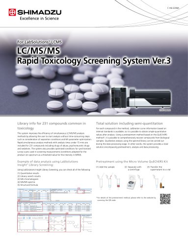 LC/MS/MS
2 Pages
 AUTOGRAPH AGX-V2 Series
28 Pages
 Metabolomics Product Portfolio
24 Pages
 Nexera UC/Nexera UC Prep
20 Pages
 Shim-pack ™ SUR-Na Columns
2 Pages
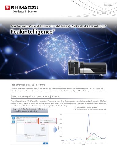 Peakintelligence
2 Pages
 Servo Controller 4830
12 Pages
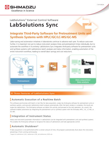 LabSolutions Sync
2 Pages
 LabSolutions Detect
8 Pages
 SALD-7500nano
16 Pages
 Multimodal Imaging System
4 Pages
 LCMS Family Brochure
12 Pages
 FluxEdge GC Systems
8 Pages
 Xslicer SMX-1010/1020
16 Pages
 LC/MS/MS MRM
2 Pages
 LC/MS/MS
2 Pages
 TRViewX
8 Pages
 UW/UX Series
4 Pages
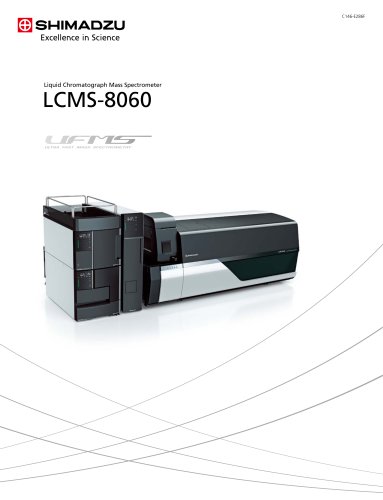 LCMS-8060
20 Pages
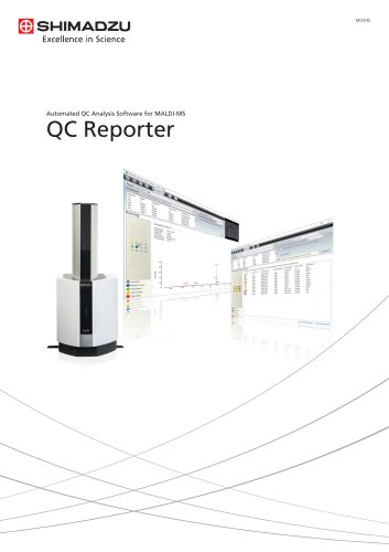 QC Reporter
6 Pages
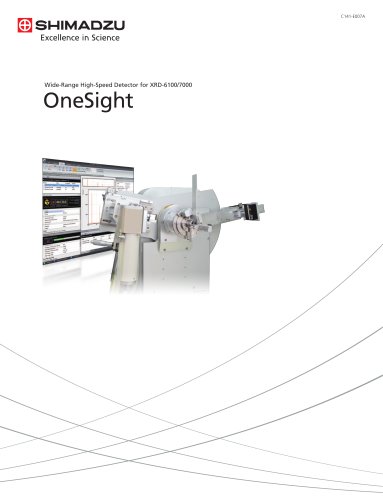 OneSight
4 Pages
 TD-30 Series
12 Pages
 Nexis GC-2030
20 Pages
 ATX/ATY series
4 Pages
 MOC63u
6 Pages
 UHPLC Nexera X2
20 Pages
 TOC-L
TOC-L16 Pages
 RF-6000
24 Pages
 ICPE-9800 series
20 Pages
 UV-3600 Plus
26 Pages
 EDX-7000/8000
24 Pages
 Capillary GC-2010 Plus
24 Pages
 GCMS QP2010 Ultra
24 Pages
 LCMS-IT-TOF
16 Pages
 Headspace Sampler HS-20
8 Pages
 BW-K/BX-K Series
2 Pages
 Catalog MOC 120H
4 Pages
 ELB SERIES
2 Pages
 TW TWC SERIES
8 Pages
 AUW-D/AUW/AUX/AUY Series
8 Pages
 Catalog UW-Series
6 Pages
 Catalog AU-Series
8 Pages
 MDGC-2010 system
1 Page





Archived catalogs
 TOC-4110
1 Page
 Catalogue HMV-2 Series
8 Pages
 Shimadzu FTIR Overview
4 Pages
 Shimadzu Software Overview
8 Pages
 Shimadzu GC/GCMS Overview
6 Pages
 Shimadzu LC/LCMS Overview
12 Pages
 Shimadzu X-ray Fluorescence
2 Pages


- Shimadzu testing machine
- Digital imager
- Visible imager
- Test cabinet
- CMOS camera module
- Industrial camera module
- Concentration analyzer
- Management software solution
- Monitoring analyzer
- Shimadzu analysis software
- Infrared imager
- Automatic analyser
- Temperature test chamber
- Real-time software
- Shimadzu material testing machine
- Shimadzu automatic testing machine
- Indentation hardness tester
- Solids analyser