- Products
- Catalogs
- News & Trends
- Exhibitions
inVia Raman microscope
1 /28Pages

inVia Raman microscope
1 /28Pages
Catalog excerpts

inVia™ research-grade confocal Raman microscopes www.renishaw.com/raman
Open the catalog to page 1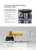
The world’s best selling high-performance Raman microscope • Designed, developed and refined over more than two decades to make it the most trusted Raman instrument on the market. • Superior, research-grade Raman microscope for your current and future needs. • Designed using Renishaw’s proven expertise in precision and innovative engineering. • Built to last: upgrade, reconfigure, or customise -the inVia Raman microscope is a sound investment. • Available in three models: inVia Basis; inVia Reflex and inVia Qontor®. • Many options and accessories are available to suit your analytical requirements...
Open the catalog to page 2
Why our users choose inVia Raman microscopes Renishaw is a global company with a worldwide network of scientists and engineers who are on-hand to provide you with expert product, technical and applications support Exceptional, reliable performance Quality, reliability and longevity The inVia system comprises a research-grade microscope coupled to a high-performance Raman spectrometer. It is simple to operate yet delivers outstanding performance—high signal throughput, combined with high spectral resolution and stability—giving reliable results, for even the most challenging measurements. The...
Open the catalog to page 3
The performance of the system, together with the excellent support from Renishaw, made the decision [to buy an inVia] an easy one for us….. inVia microscopes are an efficient, easy-to-use, easy-to-share system. Boston University (USA) inVia™ confocal Raman microscop
Open the catalog to page 4
Key benefits High performance Repeatable results The inVia Raman microscope delivers outstanding performance, providing you with the best data in the shortest time. Rely on the inVia Raman microscope to produce results you can trust. With its outstanding performance you can be confident that it will deliver repeatable results time and time again – no matter how challenging the experiment. Sensitive See even the weakest Raman scatterers and get spectra from thin films and monolayers. Powerful Use the inVia Raman microscope for both Raman and photoluminescence measurements to obtain information...
Open the catalog to page 5
Key features High optical efficiency High spectral stability Fast and sensitive analysis Get consistent, reliable data Renishaw’s engineers have used their vast experience of precision and innovative design to make the inVia Raman microscope the most sensitive Raman instrument available. They use a stigmatic on-axis spectrometer which gives highest optical efficiency, excellent stray light rejection and exceptional sensitivity. With the inVia system you can study very weak Raman signals and rapidly analyse even minute traces of material. With its rigid, lightweight baseplate, and precision kinematic...
Open the catalog to page 6
intensity / arbitrary A Raman spectrum of l-histidine, showing the lattice modes (inset), fingerprint and C-H ranges, at high spectral resolution. It was acquired in a single spectral collection using SynchroScan™ technology.
Open the catalog to page 7
The inVia Raman microscope supports a wide range of environmental and sampling accessories. Here, a mapping experiment is being performed at high temperature. Highly sensitive detectors High performance microscope Cutting edge technology Leica for quality, efficiency and reliability The inVia Raman microscope's use Renishaw’s own ultra-low noise, ultra-high sensitivity CCD cameras so you get the best results in the shortest possible time. Should you wish to add more, the inVia system can be fitted with up to four detectors, such as electron multiplied (EM) detectors and InGaAs arrays. Leica Microsystems’...
Open the catalog to page 8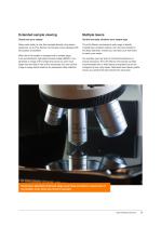
Extended sample viewing Multiple lasers Clearly see your sample Get the best data, whatever your sample type Many users prefer to view their samples directly using stereo eyepieces, so all inVia Raman microscopes come equipped with binoculoars as standard. The inVia Raman microscope’s wide range of directly coupled laser excitation options, from the near-infrared to the deep ultraviolet, ensures you can tailor your instrument to match your needs. When the inVia system is equipped with a sample stage, such as Renishaw’s high speed encoded stage (MS30), it can generate an image of the sample that...
Open the catalog to page 9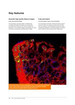
Crisp, clear chemical images The inVia Raman microscope offers a complete range of imaging technologies that enable you to acquire data from points, lines, areas, and even volumes. StreamLine™, StreamHR™ and True Raman Imaging™ technologies are unique to Renishaw and generate outstanding Raman images. The inVia system is easy to use and maintain The inVia system’s automation removes the need for manual intervention. When you change key components, like filters, lasers and gratings, it will automatically reconfigure its optics and optimise its alignment. This makes analysis more efficient, which...
Open the catalog to page 10
Maintain focus in real time Raman polarisation option Sample surface/interface tracking technology For analysis of the symmetry and orientation of samples Use LiveTrack™ automated focus-tracking technology to acquire, in real-time, accurate and repeatable spectra and topography from samples with extensive variations in height. Create stunning 3D images of uneven, curved or rough surfaces without the need for pre-scanning. Optional polarisation enables the control of both laser and spectrometer polarisation (polariser/analyser). With these options you can determine the orientation of crystals...
Open the catalog to page 11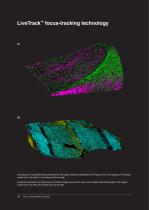
(a) Analysis of a coated flat-head screwdriver bit. The image reveals the distributions of TiN (green) and TiO2 (magenta). The imaged surface is 8.1 mm wide, 5.1 mm deep and 3.6 mm high. (b) Quartz-dominated rock (Tiger’s Eye). The Raman image shows quartz (cyan) and inorganic carbonates (yellow). The imaged surface is 47 mm wide, 26 mm deep, and 3.0 mm high. 12 inVia™ confocal Raman microscope
Open the catalog to page 12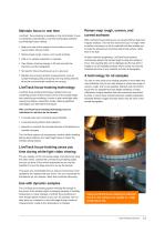
Maintain focus in real time LiveTrack focus-tracking is available on the inVia Qontor. Focus is maintained automatically in real time during data collection and white-light video viewing. ™ • Keep your view of the sample in focus while you move around under manual control. • Raman-image rough, uneven, and curved surfaces. • Little or no sample preparation is required. • View Raman chemical images in 3D and see both the chemistry and the topography. • No need for time-consuming set up or a pre-scan. • Maintain focus during dynamic measurements, such as sample heating/cooling and during very long...
Open the catalog to page 13All RENISHAW catalogs and technical brochures
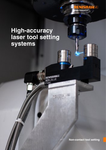
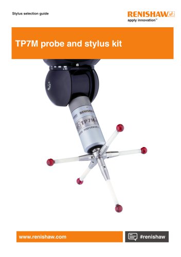 TP7M probe and stylus kit
4 Pages
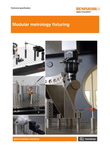 Renishaw fixtures
68 Pages
 QC20 ballbar
16 Pages
 Precision styli brochure
40 Pages
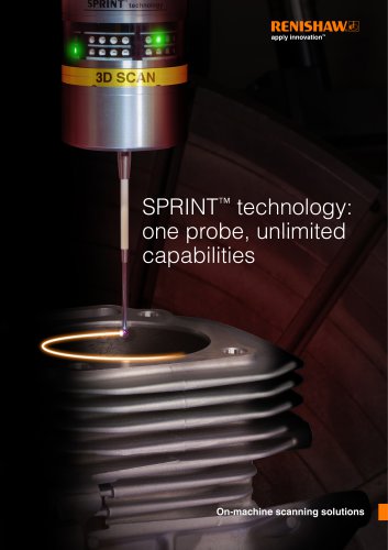 SPRINT technology brochure
11 Pages
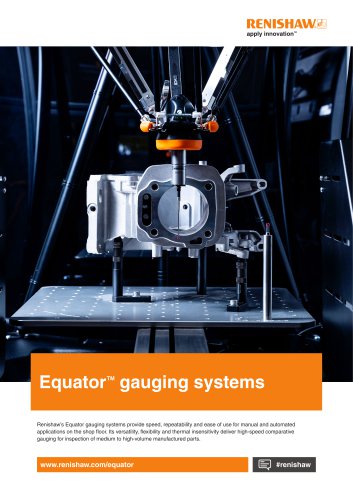 Equator brochure
12 Pages
 Metrology fixture table
4 Pages
 RFP1 fringe probe for REVO-2
2 Pages
 RVP vision probe for REVO-2
2 Pages
 MH20 articulating probe head
2 Pages
 Data sheet: MH20 and MH20i
4 Pages
 RTP20
RTP202 Pages
 PH10M-iQ PLUS
2 Pages
 REVO-2 and RSP2 probes
2 Pages
 SFP2 surface finish probe
2 Pages
 Data sheet: HS20 laser head
2 Pages
 Data sheet: RLU20 laser unit
2 Pages
 Data sheet: RLU10 laser unit
2 Pages
 RLMD01_09
13 Pages
 RLBD01_04
9 Pages
 RLCD03_03
9 Pages
 HiLin™
20 Pages
 PRIMO™ system
8 Pages
 RSP3-6 extended reach probe
4 Pages
 RGH25F UHV, RGH20F UHV
8 Pages
 Renishaw retrofit
12 Pages
 SP80
SP804 Pages
 SP600
SP6004 Pages
 Styli for Zeiss applications
57 Pages
 Precision styli
60 Pages
 CMM technology guide
28 Pages


- Temperature probe
- RENISHAW rotary encoder
- Measuring machine
- RENISHAW incremental encoder
- Calibration system
- RENISHAW incremental rotary encoder
- Monitoring software solution
- RENISHAW absolute rotary encoder
- Measurement software
- Thermocouple temperature transducer
- Solid-shaft rotary encoder
- Laboratory microscope
- Automated software
- RENISHAW optical rotary encoder
- Programming software
- RENISHAW magnetic rotary encoder
- RENISHAW industrial rotary encoder
- IP67 rotary encoder