- Company
- Products
- Catalogs
- News & Trends
- Exhibitions
TCS SP8
1 /20Pages

TCS SP8
1 /20Pages
Catalog excerpts

Location, Country, Year. Subject to modifications. LEICA and the Leica Logo are registered trademarks of Leica Microsystems IR GmbH. Order no.: English ??? ??? ∙ ??/11/???/???? ∙ Copyright © by Leica Microsystems ?????, Leica TCS SP8 – Confocal Platform
Open the catalog to page 1
“It is great if you can buy, with a small budget, a relatively standard microscope setup and if you need a special technique, you can upgrade it at a relatively low price.” “When you start with a microscope configuration for your application, you don’t know how the research will develop. Especially in imaging facilities, we need to be flexible. And this flexibility is what I really like about the system.” “I am very fond of the concept of modularity, because you don’t have to buy entirely new equipment every time you need additional capability.” Bram van de Broek Netherlands Cancer Institute,...
Open the catalog to page 3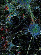
Rat cortical neurons (prim. culture). Max. projection. Nuclei (Dapi, blue), Nestin (Cy2, green), DCX (Cy3, red), ßIII-tubulin (Cy5, white)
Open the catalog to page 4
THINK MODULAR: WHERE EFFECTIVENESS MEETS EFFICIENCY The Leica TCS SP8 is designed to meet all of your future requirements with effectiveness and efficiency. >> et exactly the microscope you need. G >> Never again compromise on speed, resolution and sensitivity. >> Preserve every precious photon. >> Ugrade your system with your growing needs.
Open the catalog to page 5
THE PLATFORM THAT IS READY FOR YOUR RESEARCH GET WHAT YOU NEED NOW AND UPGRADE WHENEVER NECESSARY. Expand your possibilities Do you know which direction your research will take in the future? From live cell to quantitative imaging, from super-sensitivity to super-resolution, from multiphoton to light sheet imaging – with the Leica TCS SP8 you can always expand your possibilities. Design your future We build on a modular concept: Tailor your microscope to your current needs and upgrade with additional functionalities at any time. Your investment in a Leica TCS SP8 will pay off – now and in the...
Open the catalog to page 6
Leica TCS SP8 CARS: Label-free imaging Leica TCS SP8 SMD: Precision in single molecule detection Leica TCS SP8 DLS: The vertical turn in light sheet imaging Leica TCS SP8 MP: Discover greater depths with multiphoton imaging
Open the catalog to page 7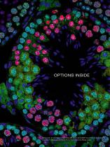
OPTIONS INSIDE Mouse testis. Synaptonemal complex (Alexa 488, green), Nuclei (DAPI, blue), Protein X (Cy3, red). Courtesy of Yu Katsuyama and Noriko Osumi, Tohoku University, Miyagi, Japan. 8
Open the catalog to page 8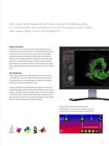
THE LAS X SOFTWARE PLATFORM CAN BE PERSONALIZED TO YOUR WORK AND ASSISTS YOU IN PLANNING, EXECUTING, AND ANALYZING YOUR EXPERIMENTS. Explore and analyze Spend your time on your research while the Leica Application Suite X (LAS X) attends to microscope control. Its workflow-oriented design and intuitive software wizards guide you step by step through image acquisition, processing, and analysis. Tailor LAS X to your needs with additional software packages like 3D Visualization and Analysis, which allow you to understand the topology of your 3D image and quantify various aspects of intracellular...
Open the catalog to page 9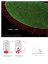
Paramecium aurelia. Epiplasmin (GFP, green), β-tubulin (Alexa 568, red) Courtesy of Anne Aubusson-Fleury, CNRS, Gif-sur-Yvette, France. Electron Bombardment Gain Avalanche Diode Avalanche Gain Principle of photomultiplier tube, which generates broad electrical pulses. Princple of hybrid detector, which results in sharp electrical pulses. Pulse Width
Open the catalog to page 10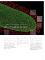
ALL OPTICAL COMPONENTS OF THE LEICA TCS SP8 ARE MATCHED TO PRESERVE YOUR PRECIOUS SIGNAL FOR BRILLIANT IMAGES FULL OF CONTRAST. More details With the synergies of the multiband spectral detector, acousto-optical beam splitter (AOBS), and super-sensitive Leica HyD hybrid detectors you get a confocal microscope with maximum photon efficiency and gapless spectral detection. The high signal-to-noise ratio of the Leica HyDs helps to render the finest details of any specimen. The superior sensitivity of the Leica TCS SP8 directly translates into less laser power and improved viability of live specimens....
Open the catalog to page 11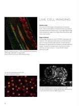
LIVE CELL IMAGING Reliable results With a Leica TCS SP8, imaging of living specimens can have many facets. It gives you the freedom to conveniently perform any advanced live imaging method with reliable results. You can expect highest image fidelity and specimen viability from its light efficient detection and high speed scanning system. Bigger and deeper Thinking bigger and deeper the Leica TCS SP8 MP accommodates live organisms. At the opposite end of the scale you can study nanoscopic detail in live cells using STED super-resolution. Or follow embryonic development using gentle light sheet...
Open the catalog to page 12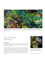
NIH3T3 cells transduced with five individual fluorescent protein (FP) vectors. FPs: Cerulean, EGFP, Venus, tdTomato and mCherry. Each FP was visible only in the cells transduced with the corresponding vector. AOBS fast line sequential scan with five excitations and five emission bands. No unmixing. Image courtesy of Daniela Malide, NIH Bethesda, MD USA MULTICOLOR IMAGING Spectral freedom Stop worrying about challenging multicolor experiments. Get the spectral freedom you need to image any kind of dye combination. Easily explore cell connectomics using Brainbow fluorescent proteins with the Leica...
Open the catalog to page 13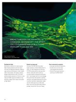
BREAK THROUGH THE BARRIERS OF CONFOCAL MICROSCOPY AND STUDY CELL DYNAMICS AND STRUCTURES SMALLER THAN 200 NM. High-resolution imaging Confocal (left) vs. Hyvolution (right) Pushing the limits Pushing your confocal to its limits requires highly sensitive detection with low noise. The Leica TCS SP8 plus HyVolution combines super-sensitive HyD detectors, which reliably collect weak signals, with industry-leading Huygens deconvolution by SVI. This combination achieves resolution of 140 nm in a case study with DNA origamis and delivers crisp multicolor images, which convey every detail at high fidelity....
Open the catalog to page 14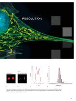
Intensity Intensity Distance/nm Distance/nm High-resolution imaging with super-sensitive HyD detectors and Huygens deconvolution can resolve 140 nm in a case study with DNA origamis. Specimen: single molecule DNA-origamis with 140 nm defined spacing. (A) Confocal image. (B) HyVolution (C) Distance measured in B (green line). (D) Distribution of measured distances in nanometers.
Open the catalog to page 15All Leica Microsystems GmbH catalogs and technical brochures
 Visoria M flyer
2 Pages
 Visoria M brochure
12 Pages
 Mateo FL Flyer EN
2 Pages
 DVM6 Brochure en
16 Pages
 Leica M50, M60, M80
12 Pages
 XL Stand
4 Pages
 LED1000
16 Pages
 LED3000 BLI
20 Pages
 LED5000 NVI
20 Pages
 LED3000 NVI
20 Pages
 LED3000 DI
20 Pages
 LED5000 HDI
20 Pages
 LED5000 CXI
20 Pages
 LED2500
8 Pages
 LED5000 MCI
20 Pages
 LED3000 MCI
20 Pages
 LED5000 SLI
20 Pages
 LED3000 SLI
20 Pages
 LED2000
8 Pages
 LED5000 RL
20 Pages
 LED3000 RL
20 Pages
 EL6000
4 Pages
 M320 F12 for ENT
12 Pages
 M620 TTS
4 Pages
 M220 F12
8 Pages
 RUV800
4 Pages
 DI C800
2 Pages
 M620 F20
8 Pages
 M822 F40 / F20
20 Pages
 M844 F40 / F20
16 Pages
 Proveo 8
16 Pages
 Envisu
8 Pages
 EnFocus
8 Pages
 M525 F20
12 Pages
 FL400
FL4004 Pages
 Leica M525 OH4
20 Pages
 Leica M720 OH5
24 Pages
 Leica M530 OH6
16 Pages
 Leica M530 OHX
16 Pages
 Leica CaptiView
4 Pages
 Leica GLOW800
4 Pages
 Leica ARveo
16 Pages
 Leica PROvido
8 Pages
 LAS X Steel Expert
4 Pages
 Leica LMD Software
16 Pages
 Leica Map
12 Pages
 Leica Cleanliness Expert
4 Pages
 Leica LAS X Industry
4 Pages
 Leica LAS X Life Science
4 Pages
 Leica EM CPD300
12 Pages
 Leica EM TP
4 Pages
 Leica EM VCT500
2 Pages
 Leica EM ACE900
12 Pages
 Leica EM ACE200
12 Pages
 Leica EM KMR3
8 Pages
 Leica EM AFS2
8 Pages
 Leica EM ICE
12 Pages
 TCS SP8 DLS
16 Pages
 Leica LIGHTNING
2 Pages
 Leica SP8 DIVE
20 Pages
 Leica K5
2 Pages
 Leica DFC295
6 Pages
 Leica DFC3000 G
6 Pages
 Leica EC4
4 Pages
 Leica DFC7000 T / DFC7000 GT
4 Pages
 Leica DFC9000
2 Pages
 Leica MZ10 F
4 Pages
 Leica M165 FC
16 Pages
 Leica DM IL LED
12 Pages
 Leica DMi1
2 Pages
 THUNDER Imager Tissue
2 Pages
 Leica FS M
12 Pages
 Leica FS C
20 Pages
 Leica FS CB
20 Pages
 Leica DM3000 / DM3000 LED
16 Pages
 Leica DM750
12 Pages
 Leica DM500
12 Pages
 Leica DM300
8 Pages
 Leica DM1750 M
12 Pages
 Leica FS4000 LED
20 Pages
 Leica DM2000 / DM2000 LED
16 Pages
 Leica DM1000
16 Pages
 Leica DM1000 LED
16 Pages
 Leica DM2500 & DM2500 LED
16 Pages
 Leica DM4 B & DM6 B
16 Pages
 Leica DMC2900
6 Pages
 Leica DMC6200
8 Pages
 Leica DMC5400
4 Pages
 Leica Z6 APO
16 Pages
 Leica Z16 APO
16 Pages
 Leica M205 FCA / M205 FA
16 Pages
 Leica DM12000 M
8 Pages
 Leica DM8000 M
8 Pages
 Leica DM4 P, DM2700 P, DM750 P
12 Pages
 Leica EM ACE600
12 Pages
 Leica EM RAPID
8 Pages
 Leica EM TRIM2
8 Pages
 Leica EM TIC 3X
16 Pages
 Leica EM TXP
10 Pages
 Leica DM2700 M
12 Pages
 DMi8 M / C / A
12 Pages
 Leica A60 S / Leica A60 F
16 Pages
 SOLUTIONS FOR MATERIALS SCIENCE
12 Pages
 Leica StereoZoom line
20 Pages
 TL-Bases
12 Pages
 TCS SP8 STED 3X
24 Pages
 TCS SP8 Objective
24 Pages
 AOBS
AOBS16 Pages
 Leica DM3 XL
7 Pages
 Leica DMi8 S
12 Pages
 Leica M50/M60/M80
12 Pages
 Leica DM750 M
12 Pages
 Leica DM4 M/ DM6 M
12 Pages
 Leica A60 S/A60 F
16 Pages


- Digital imager
- Visible imager
- CMOS camera module
- Industrial camera module
- Nora LED lighting
- Nora analysis software
- Positioning table
- Full-color camera system
- Nora Windows software
- Translation stage
- USB camera module
- Monochrome camera module
- 3D software solution
- Nora industrial software
- Nora optical microscope
- HD camera module
- LED hand lamp
- Quality software
- Nora laboratory microscope