 Website:
Leica Microsystems GmbH
Group: DANAHER
Catalog excerpts
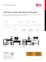
A Decade of Super-Resolution Innovation Working with passion for the global research community: The TCS SP8 STED 3X marks one decade of innovative super-resolution technology by Leica Microsystems - providing quality data and efficient processes for your science. TCS STED Dual color for TCS SP8 STED TCS SP8 TCS STED (CW) and gated STED STED 3X CONNECT WITH US LASER RADIATION VISIBLE AND INVISIBLE - CLASS 4 AVOID EYE OR SKIN EXPOSURE TO DIRECT OR SCAITERED RAOIATION P< 4W 350-1600nm >801 s IEC 60825-1 : 2007 www.leica-microsystems.com/ products/super-resolution Order no.: English 1593003011 • Copyright © by Leica Microsystems CMS GmbH, Mannheim, Germany, 2014. Subject to modifications. LEICA and the Leica Logo are registered trademarks of Leica Microsystems IR GmbH.
Open the catalog to page 1
Leica TCS SP8 STED 3X Your Next Dimension!
Open the catalog to page 2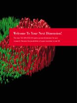
Welcome To Your Next Dimension! The new TCS SP8 STED 3X opens up new dimensions for your research: Discover the possibilities of super-resolution in real 3D.
Open the catalog to page 4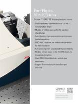
Pure Physics, More Efficiency The new TCS SP8 STED 3X strengthens your science: ››Tunable and direct super-resolution in x, y and z reveal smallest details ›› Multiple STED lines open up the full spectrum of visible light ›› Gated detection improves resolution and increases live cell capabilities ›› STED WHITE objective has optimal color correction for the full spectrum ›› Auto beam alignment provides stability and reliability ›› Modular concept based on the TCS SP8 allows you to upgrade at any time ›› Smart STED Wizard intuitively controls your experiments ›› Huygens deconvolution gets...
Open the catalog to page 6
TCS SP8 STED 3X – Here’s Your Next Dimension! Here’s Your Next Dimension! STED microscopy by Leica Microsystems has revolutionized the study of subcellular a rchitecture and cell dynamics at the nanoscale. The fully integrated STED (STimulated Emission Depletion) system meets the requirements of daily research and provides fast, intuitive, and purely optical access to structural details far beyond the diffraction limit. Gated STED substantially improves resolution to below 50 nm and increases live cell viability. Now, the next generation of STED, the TCS SP8 STED 3X, broadens the scope of...
Open the catalog to page 8
Pure Physics, More Reliability STimulated Emission Depletion imaging reaches lateral resolution below 50 nm in true optical sections by downscaling the spot from where fluorescence is generated. STED is pure physics: What you see is what you get. The principle Super-resolution – fast and direct The basic principle of STED, which was A phase mask filter determines the area STED technology provides fast and direct first described by Stefan Hell in 1994 , is in the focus plane where STED light is access to structural details at the nano- simple. The effective focal spot scanning dominant, e.g....
Open the catalog to page 9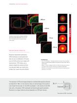
TCS SP8 STED 3X – Pure Physics, More Reliability STED Intensity Achieved resolution (left) correlates with STED laser power (right). Right panel: STED donut with area of stimulated emission in red, area of fluorescence in green. Resolution improvement achieved by STED microscopy is purely optical and does not rely on mathematics. Of course, you can further process your images with deconvolution. STED allows you to directly compare the outcome with the raw data and makes your results more reliable. Don’t waste time with artifacts! Key Publications ›› Hell, S. W. & Wichmann, J. Breaking the...
Open the catalog to page 10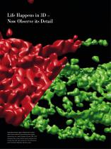
Life Happens in 3D – Now Observe its Detail Immunofluorescence stain of Golgi marker in HeLa cells: Surface rendered 3D reconstruction after Huygens deconvolution of a z stack (20 planes), 3D donut 100%. STED data (green) show a clear resolution increase compared to the confocal result (red). Data courtesy of Dr. Timo Zimmermann, Center for Genomic Regulation, Barcelona, Spain.
Open the catalog to page 11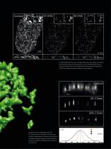
Histone H3-Alexa 568 in HeLa cells: The highest lateral resolution increase is achieved by the vortex donut, maximal resolution increase in x, y and z by 3D STED. Note the loss of structures of objects that are not in the super-resolved focus plane when using 3D STED. 3D STED also achieves an increase of resolution in the xy dimension. 11#ii itx axisz axis0% Z Donut60% Z Donut XZ axis plot of Histone H3-Alexa 568 in HeLa cells. 0% z donut allocates all STED light to the vortex donut resulting in maximal resolution increase in x and y, but not in z compared to confocal microscopy. A small...
Open the catalog to page 12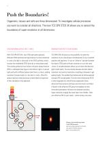
Push the Boundaries! Organisms, tissues and cells are three dimensional. To investigate cellular processes you want to consider all directions. The new TCS SP8 STED 3X allows you to extend the boundaries of super-resolution in all dimensions. Discover more details in X, Y and Z Engineer your PSF to your science With TCS SP8 STED 3X, two STED light paths generate TCS SP8 STED 3X gives you the possibility to match the different STED patterns (see figure below). For best resolution resolution of your microscope in all dimensions to your scientific in x and y the light is allocated to the STED...
Open the catalog to page 13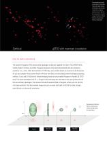
Immunostaining of endosomal marker (Lamp M) in TCS SP8 STED 3X – Subchapter 13 HeLa cells. Note the loss of structures out of superresolved focal plane (white arrows) in the middle compared to confocal images. Courtesy of Shem Johnson, University of Geneva, Switzerland. gSTED with maximum z resolution Push the limits even further The powerful Huygens STED deconvolution package, exclusively supplied with every TCS SP8 STED 3X system, helps to improve your data. Huygens decreases noise levels dramatically and also enhances resolution in x, y and z. After deconvolution of STED data, even...
Open the catalog to page 14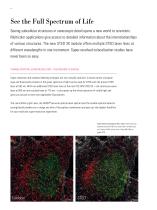
See the Full Spectrum of Life Seeing subcellular structures in nanoscopic detail opens a new world to scientists. Multicolor applications give access to detailed information about the interrelationships of various structures. The new STED 3X module offers multiple STED laser lines at different wavelengths in one instrument. Super-resolved colocalization studies have never been so easy. Tunable spectral super-resolution – the freedom to choose Super-resolution and standard labeling strategies are not mutually exclusive. A broad variety of popular dyes and fluorescent proteins in the green...
Open the catalog to page 15All Leica Microsystems GmbH catalogs and technical brochures
-
Mateo FL Flyer EN
2 Pages
-
DVM6 Brochure en
16 Pages
-
Leica M50, M60, M80
12 Pages
-
XL Stand
4 Pages
-
LED1000
16 Pages
-
LED3000 BLI
20 Pages
-
LED5000 NVI
20 Pages
-
LED3000 NVI
20 Pages
-
LED3000 DI
20 Pages
-
LED5000 HDI
20 Pages
-
LED5000 CXI
20 Pages
-
LED2500
8 Pages
-
LED5000 MCI
20 Pages
-
LED3000 MCI
20 Pages
-
LED5000 SLI
20 Pages
-
LED3000 SLI
20 Pages
-
LED2000
8 Pages
-
LED5000 RL
20 Pages
-
LED3000 RL
20 Pages
-
EL6000
4 Pages
-
M320 F12 for ENT
12 Pages
-
M620 TTS
4 Pages
-
M220 F12
8 Pages
-
RUV800
4 Pages
-
DI C800
2 Pages
-
M620 F20
8 Pages
-
M822 F40 / F20
20 Pages
-
M844 F40 / F20
16 Pages
-
Proveo 8
16 Pages
-
Envisu
8 Pages
-
EnFocus
8 Pages
-
M525 F20
12 Pages
-
FL400
4 Pages
-
Leica M525 OH4
20 Pages
-
Leica M720 OH5
24 Pages
-
Leica M530 OH6
16 Pages
-
Leica M530 OHX
16 Pages
-
Leica CaptiView
4 Pages
-
Leica GLOW800
4 Pages
-
Leica ARveo
16 Pages
-
Leica PROvido
8 Pages
-
LAS X Steel Expert
4 Pages
-
Leica LMD Software
16 Pages
-
Leica Map
12 Pages
-
Leica Cleanliness Expert
4 Pages
-
Leica LAS X Industry
4 Pages
-
Leica LAS X Life Science
4 Pages
-
Leica EM CPD300
12 Pages
-
Leica EM TP
4 Pages
-
Leica EM VCT500
2 Pages
-
Leica EM ACE900
12 Pages
-
Leica EM ACE200
12 Pages
-
Leica EM KMR3
8 Pages
-
Leica EM AFS2
8 Pages
-
Leica EM ICE
12 Pages
-
TCS SP8 DLS
16 Pages
-
Leica LIGHTNING
2 Pages
-
Leica SP8 DIVE
20 Pages
-
Leica K5
2 Pages
-
Leica DFC295
6 Pages
-
Leica DFC3000 G
6 Pages
-
Leica EC4
4 Pages
-
Leica DFC7000 T / DFC7000 GT
4 Pages
-
Leica DFC9000
2 Pages
-
Leica MZ10 F
4 Pages
-
Leica M165 FC
16 Pages
-
Leica DM IL LED
12 Pages
-
Leica DMi1
2 Pages
-
THUNDER Imager Tissue
2 Pages
-
Leica FS M
12 Pages
-
Leica FS C
20 Pages
-
Leica FS CB
20 Pages
-
Leica DM3000 / DM3000 LED
16 Pages
-
Leica DM750
12 Pages
-
Leica DM500
12 Pages
-
Leica DM300
8 Pages
-
Leica DM1750 M
12 Pages
-
Leica FS4000 LED
20 Pages
-
Leica DM2000 / DM2000 LED
16 Pages
-
Leica DM1000
16 Pages
-
Leica DM1000 LED
16 Pages
-
Leica DM2500 & DM2500 LED
16 Pages
-
Leica DM4 B & DM6 B
16 Pages
-
Leica DMC2900
6 Pages
-
Leica DMC6200
8 Pages
-
Leica DMC5400
4 Pages
-
Leica Z6 APO
16 Pages
-
Leica Z16 APO
16 Pages
-
Leica M205 FCA / M205 FA
16 Pages
-
Leica DM12000 M
8 Pages
-
Leica DM8000 M
8 Pages
-
Leica DM4 P, DM2700 P, DM750 P
12 Pages
-
Leica EM ACE600
12 Pages
-
Leica EM RAPID
8 Pages
-
Leica EM TRIM2
8 Pages
-
Leica EM TIC 3X
16 Pages
-
Leica EM TXP
10 Pages
-
Leica DM2700 M
12 Pages
-
DMi8 M / C / A
12 Pages
-
Leica A60 S / Leica A60 F
16 Pages
-
SOLUTIONS FOR MATERIALS SCIENCE
12 Pages
-
Leica StereoZoom line
20 Pages
-
TL-Bases
12 Pages
-
TCS SP8
20 Pages
-
TCS SP8 Objective
24 Pages
-
AOBS
16 Pages
-
Leica DM3 XL
7 Pages
-
Leica DMi8 S
12 Pages
-
Leica M50/M60/M80
12 Pages
-
Leica DM750 M
12 Pages
-
Leica DM4 M/ DM6 M
12 Pages
-
Leica A60 S/A60 F
16 Pages


