- Catalogs
- Leica Microsystems GmbH
- Leica SP8 DIVE
- Company
- Products
- Catalogs
- News & Trends
- Exhibitions
Leica SP8 DIVE
1 /20Pages

Leica SP8 DIVE
1 /20Pages
Catalog excerpts

SP8 DIVE — DEEP IN VIVO EXPLORER The world’s first tunable deep imaging solution
Open the catalog to page 1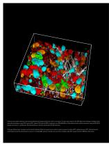
Confetti mouse small intestines, fluorescently labeled and lineage traced from a multi-color tracer. The gray color codes for the SHG signal from Collagen. Lineage traced stem cells are shown in cyan (CFP), green (GFP), yellow (YFP) and red (RFP). Image size is ca. 700x700x150 µm³. Recorded with two-photon excitation, using the SP8 DIVE. Sample courtesy of J. van Rheenen, Netherlands Cancer Institute, Amsterdam (the Netherlands). Title page: Mouse brain, transgenic and transiently labeled. Different types of nerve cells are shown in green (microglia, GFP), yellow (neurons, YFP), blue (astrocytes,...
Open the catalog to page 2
SP8 DIVE SPECTRAL FREEDOM FOR MULTICOLOR DEEP IN VIVO IMAGING Spectral Freedom with 4Tune Up to now, the possibilities to do multicolor deep in vivo experiments were limited by the choice of dichroic filters. The SP8 with the brand new and unique 4Tune detector is the world’s first fully spectrally tunable multiphoton microscope. Another degree of spectral freedom results from the newly implemented possibility to use up to three IR excitation lines. Today’s research focuses on complex biological processes such as human diseases. Complexity mainly results from the number of components involved:...
Open the catalog to page 3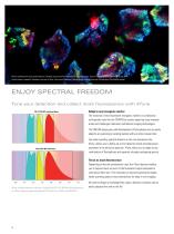
4Tune enables new dye combinations: Confetti mouse small intestine. Cell lineage tracer. Cyan: CFP, green: GFP, yellow: YFP, red: RFP. Colon cancer research. Sample courtesy of Prof. Jacco van Rheenen, Netherlands Cancer Institute, Amsterdam (the Netherlands). ENJOY SPECTRAL FREEDOM Tune your detection and collect more fluorescence with 4Tune Adapt to any transgenic marker The evolution of new fluorescent transgenic markers in combination with genetic tools like the CRISPR/Cas system speed up many research areas and challenges traditional multiphoton imaging technologies. CFP, YFP & RFP with...
Open the catalog to page 4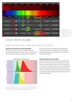
♦ Cooled Standard * MP-EYFPi I The intuitive 4Tune user interface of the Leica LAS X software. Up to four channels can be defined and imaged simultaneously - unlimited in the sequential mode. DIVE WITH EASEExperimental set-up made easy with Leica LAS X Optimize your settings live and with minimal effort The intuitive 4Tune user interface lets you optimize the emission settings of multiple fluorophores live and on the fly. Due to its clear and user-friendly design, operation is easy and requires minimal training. If more than four markers are investigated or overlapping spectra require sequential...
Open the catalog to page 5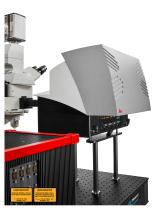
LASER RADIATION LASER RADIATION LASER RADIATION LASER RADIATION VISIBLE VISIBLE AND INVISIBLE-3B AND INVISIBLE- CLASS CLASS 3B AVOID DIRECT EXPOSURE TO BEAM BEAM AVOID DIRECT EXPOSURE TO VISIBLE VISIBLE AND INVISIBLE-4CLASS 4 AND INVISIBLE- CLASS AVOID EYE OR SKINOR SKIN EXPOSURE TO AVOID EYE EXPOSURE TO DIRECT OR SCATTERED RADIATION DIRECT OR SCATTERED RADIATION
Open the catalog to page 6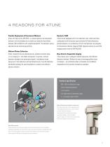
4 REASONS FOR 4TUNE Flexible Deployment of Fluorescent Markers 4Tune, the heart of the SP8 DIVE, is a unique spectral non-descanned detection unit that enables you to define your detection band freely between 380 and 800 nm with variable bandwidth. The detection can be adjusted live by simple drag and drop. Efficient Photon Collection 4Tune, located at the non-descanned site, enables multicolor deep in vivo imaging in 1 mm depth and beyond. In contrast, confocal detection strongly limits penetration depth in multiphoton mode. Equip your 4 Tune detection with HyD detection and tune the detection...
Open the catalog to page 7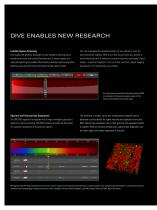
DIVE ENABLES NEW RESEARCH Lambda Square Scanning Fully explore the photonic landscape of your sample by carrying out an excitation-emission scan using 4Tune detection. Emission spectra are captured exploiting the tunable 4Tune bands, excitation spectra acquisition becomes easy using the newly introduced constant power mode. You can investigate the autofluorescence of your sample or hunt for new fluorescent markers. With only a few mouse-clicks you receive a two-dimensional plot of excitation versus emission for each pixel of your image – a spectral fingerprint. You can either use this to adjust...
Open the catalog to page 8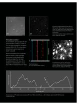
Comparison of 2PE with 920 nm (left) and 3PE with 1300 nm (right), 800 pm deep in the live mouse brain cortex. Thyl-eYFP cortical neurons, IRAPO 25x1.0 W motCorr, both images autoscaled from darkest to brightest pixel. Sample courtesy of Kevin Keppler, Light Microscope Facility, DZNE, Bonn (Germany). Simplified principles of one-, two - and three-photon excitation. Three photon excitation The new SP8 DIVE can be equipped with laser sources that allow excitation up to 1300 nm. This is the ideal wavelength for three-photon excitation (3PE) of GFP, YFP or other markers within the same spectral range...
Open the catalog to page 9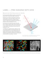
LABEL — FREE IMAGING WITH DIVE Second and third harmonics on the fly Second harmonic generation (SHG) has emerged as a powerful label-free imaging modality to visualize fibrillar collagen in diverse tissues. SHG signals occur from large non-centrosymmetric structures with a periodic alignment. These structures double the frequency of the infrared pulsed excitation light used for multiphoton imaging. In conclusion, no labels need to be incorporated and since the light is not absorbed but scattered, photodamage does not occur. The information from this channel can be used in various ways: Either...
Open the catalog to page 10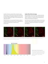
Like SHG, third harmonic generation (THG) is a label-free imaging method, however it occurs at exactly one third of the incoming pulsed laser light wavelength. By equipping the SP8 DIVE with a laser source that provides wavelengths up to 1300 nm, THG can be detected in the visible range at 430 nm. Combine THG with Ultra-fast imaging Blood flow happens at very fast time scales. To enable live blood flow tracking, the optional 12 kHz resonant scanner with frame rates up to 428 fps, is the ideal tool. In combination with the ultrasensitive HyD detection even weak signals can be picked up. With 4Tune...
Open the catalog to page 11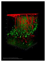
Mouse brain cortex., Thy1-eYFP, Tx Red. IRAPO 25x1.0 W motCorr. Sample courtesy of Kevin Keppler, Light Microscope Facility, DZNE Bonn (Germany).
Open the catalog to page 12All Leica Microsystems GmbH catalogs and technical brochures
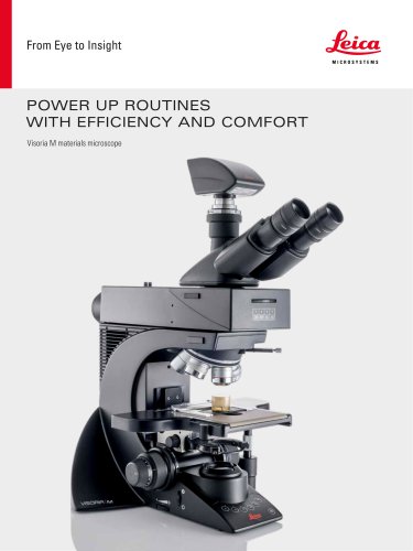
 Visoria M flyer
2 Pages
 Visoria M brochure
12 Pages
 Mateo FL Flyer EN
2 Pages
 DVM6 Brochure en
16 Pages
 Leica M50, M60, M80
12 Pages
 XL Stand
4 Pages
 LED1000
16 Pages
 LED3000 BLI
20 Pages
 LED5000 NVI
20 Pages
 LED3000 NVI
20 Pages
 LED3000 DI
20 Pages
 LED5000 HDI
20 Pages
 LED5000 CXI
20 Pages
 LED2500
8 Pages
 LED5000 MCI
20 Pages
 LED3000 MCI
20 Pages
 LED5000 SLI
20 Pages
 LED3000 SLI
20 Pages
 LED2000
8 Pages
 LED5000 RL
20 Pages
 LED3000 RL
20 Pages
 EL6000
4 Pages
 M320 F12 for ENT
12 Pages
 M620 TTS
4 Pages
 M220 F12
8 Pages
 RUV800
4 Pages
 DI C800
2 Pages
 M620 F20
8 Pages
 M822 F40 / F20
20 Pages
 M844 F40 / F20
16 Pages
 Proveo 8
16 Pages
 Envisu
8 Pages
 EnFocus
8 Pages
 M525 F20
12 Pages
 FL400
FL4004 Pages
 Leica M525 OH4
20 Pages
 Leica M720 OH5
24 Pages
 Leica M530 OH6
16 Pages
 Leica M530 OHX
16 Pages
 Leica CaptiView
4 Pages
 Leica GLOW800
4 Pages
 Leica ARveo
16 Pages
 Leica PROvido
8 Pages
 LAS X Steel Expert
4 Pages
 Leica LMD Software
16 Pages
 Leica Map
12 Pages
 Leica Cleanliness Expert
4 Pages
 Leica LAS X Industry
4 Pages
 Leica LAS X Life Science
4 Pages
 Leica EM CPD300
12 Pages
 Leica EM TP
4 Pages
 Leica EM VCT500
2 Pages
 Leica EM ACE900
12 Pages
 Leica EM ACE200
12 Pages
 Leica EM KMR3
8 Pages
 Leica EM AFS2
8 Pages
 Leica EM ICE
12 Pages
 TCS SP8 DLS
16 Pages
 Leica LIGHTNING
2 Pages
 Leica K5
2 Pages
 Leica DFC295
6 Pages
 Leica DFC3000 G
6 Pages
 Leica EC4
4 Pages
 Leica DFC7000 T / DFC7000 GT
4 Pages
 Leica DFC9000
2 Pages
 Leica MZ10 F
4 Pages
 Leica M165 FC
16 Pages
 Leica DM IL LED
12 Pages
 Leica DMi1
2 Pages
 THUNDER Imager Tissue
2 Pages
 Leica FS M
12 Pages
 Leica FS C
20 Pages
 Leica FS CB
20 Pages
 Leica DM3000 / DM3000 LED
16 Pages
 Leica DM750
12 Pages
 Leica DM500
12 Pages
 Leica DM300
8 Pages
 Leica DM1750 M
12 Pages
 Leica FS4000 LED
20 Pages
 Leica DM2000 / DM2000 LED
16 Pages
 Leica DM1000
16 Pages
 Leica DM1000 LED
16 Pages
 Leica DM2500 & DM2500 LED
16 Pages
 Leica DM4 B & DM6 B
16 Pages
 Leica DMC2900
6 Pages
 Leica DMC6200
8 Pages
 Leica DMC5400
4 Pages
 Leica Z6 APO
16 Pages
 Leica Z16 APO
16 Pages
 Leica M205 FCA / M205 FA
16 Pages
 Leica DM12000 M
8 Pages
 Leica DM8000 M
8 Pages
 Leica DM4 P, DM2700 P, DM750 P
12 Pages
 Leica EM ACE600
12 Pages
 Leica EM RAPID
8 Pages
 Leica EM TRIM2
8 Pages
 Leica EM TIC 3X
16 Pages
 Leica EM TXP
10 Pages
 Leica DM2700 M
12 Pages
 DMi8 M / C / A
12 Pages
 Leica A60 S / Leica A60 F
16 Pages
 SOLUTIONS FOR MATERIALS SCIENCE
12 Pages
 Leica StereoZoom line
20 Pages
 TL-Bases
12 Pages
 TCS SP8
20 Pages
 TCS SP8 STED 3X
24 Pages
 TCS SP8 Objective
24 Pages
 AOBS
AOBS16 Pages
 Leica DM3 XL
7 Pages
 Leica DMi8 S
12 Pages
 Leica M50/M60/M80
12 Pages
 Leica DM750 M
12 Pages
 Leica DM4 M/ DM6 M
12 Pages
 Leica A60 S/A60 F
16 Pages


- Digital imager
- Visible imager
- CMOS camera module
- Industrial camera module
- Nora LED lighting
- Nora analysis software
- Positioning table
- Full-color camera system
- Nora Windows software
- Translation stage
- USB camera module
- Monochrome camera module
- 3D software solution
- Nora industrial software
- Nora optical microscope
- HD camera module
- LED hand lamp
- Quality software
- Nora laboratory microscope