- Catalogs
- Leica Microsystems GmbH
- Leica M525 OH4
- Company
- Products
- Catalogs
- News & Trends
- Exhibitions
Leica M525 OH4
1 /20Pages

Leica M525 OH4
1 /20Pages
Catalog excerpts

M525 OH4 Surgical Microscope Solution for Precision Neurosurgery
Open the catalog to page 1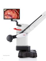
OH4 stand designed by Mitaka
Open the catalog to page 2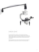
M525 OH4 Leica Microsystems stands for excellence in optics. Outstanding contrast, brilliance, sharpness, resolution, color fidelity, and precision are hallmarks of surgical microscopes. The OH4 stand, designed by Mitaka, not only complements the M525 optics, but also improves the overall microsurgical experience with superior movement, innovative illumination, and userfriendly features. Designed and manufactured using superior materials and the highest quality standards, the premium M525 OH4 miscroscope is built for long service life and outstanding reliability.
Open the catalog to page 3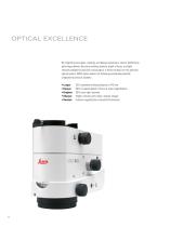
OPTICAL EXCELLENCE By integrating new glass, coatings, and design parameters, Leica’s OptiChrome technology delivers the extra working distance, depth of focus, and light intensity needed for precision microsurgery. It forms the basis for the premium optical system. M525 optics deliver the following out tanding benefits s compared to previous systems: • Longer • Deeper • Brighter • Sharper • Smarter 32% extended working distance to 470 mm 30% increased depth of focus at same magnification 30% more light intensity Higher contrast and crisper, sharper images AutoIris magnification-controlled il
Open the catalog to page 4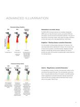
ADVANCED ILLUMINATION Illumination Settings: BrightCare Max BrightCare Adjusted Illumination in an instant, always Working Distance The M525 OH4 microscope features two completely independent 300W xenon arc-lamp illumination systems, plus optional 400-watt illumination. The second system auto atically activates in the event of m lamp failure in the primary system, which gives the surgeon confidence to know that surgery will not be jeopardized due to lamp failure. BrightCare – Working-distance-controlled illumination working distance working distance auto adjustment for safer illumination levels...
Open the catalog to page 5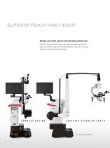
SUPERIOR REACH AND HEIGHT Compact, yet provides superior reach and ample overhead room The M525 OH4 microscope features high overhead clearance and superior reach, providing the surgeon with ultimate flexibility to place the microscope wherever is most beneficial for the surgery. OH4 stand designed by Mitaka
Open the catalog to page 6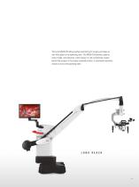
The Leica M525 OH4 allows perfect positioning for surgery and takes up very little space in the operating room. The M525 OH4 provides superior reach, height, and clearance, which allows it to be conveniently located behind the surgeon n the unique overhead position, or positioned anywhere i around or across the operating table.
Open the catalog to page 7
WIDE RANGE OF MOVEMENT Extraordinary movement The M525 OH4 microscope offers a wide range of movement in all dimensions for improved maneuverability. The microscope can be moved with very little force, and has minimal vibration at all magnification levels. The stand’s patented advanced movement system achieves perfect balance in six axes and at all locations and angles of the microscope. 100° range of lateral movement provides the most difficult-tomaneuver side views. 150° inclination angle range combined with the most compact microscope provides unmatched comfort, even in difficult positions....
Open the catalog to page 8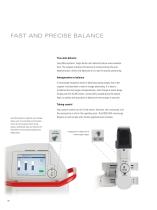
FAST AND PRECISE BALANCE True auto-balance Leica Microsystems’ single button auto balance feature saves valuable time. The surgeon activates this feature by simply pushng the autoi balance button, which fully balances all six axes for precise positioning. Intraoperative re-balance A microscope frequently needs re-balancing during surgery due to the surgeon’s and assistant’s need to change positioning. It is easy to re-balance the microscope intraoperatively, even through a sterile drape. Simply push the AC/BC button, conveniently located above the optical head, to quickly and accurately re-balance...
Open the catalog to page 10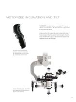
MOTORIZED INCLINATION AND TILT The M525 OH4 has robotic functions on two axes (X/Y) to further enhance movement precision. The robotic functions can be activated by hand and/or foot controls. Combined with an IGS computer, the stand’s robotic ability allows the microscope to follow the surgeon’s hand instruments, thus eliminating the need for the surgeon to take his/her hand away from the surgical site to move the microscope. The handle’s ultra precise joystick allows micrometric movements for tilt and inclination. When preselected, it can also, for example, control the image injection functions....
Open the catalog to page 11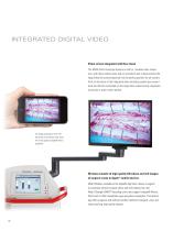
INTEGRATED DIGITAL VIDEO Video screen integrated with floor stand The M525 OH4 microscope features a built-in, movable video screen arm, with three rotation axes and an inclination axis to best position the large video flat screen (optional) into the perfect position for all viewers. Also, all functions of the integrated video recording system are conveniently and directly controlled via the large video screen (using a keyboard, touch pad or touch screen option). Full image coverage on a 24” HD flat screen and wireless video transfer to the surgeon’s Apple® device (optional). Wireless transfer...
Open the catalog to page 12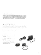
Ready for future imaging technologies The selection of video options continually changes as imaging technology evolves. The M525 OH4 is an open architecture system that allows the surgeon to upgrade components as new video innovations become available. The Med X Change HDMD® high-definition digital recording system is easily integrated with the Leica M525 OH4 floor stand for convenience and easy accessibility. Wide choice of Leica Video Adapters All Leica video adapters offer an intra operative fine focus to adjust the video focus. This enables the surgeon to always achieve crisp and clear focus...
Open the catalog to page 13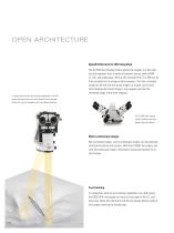
OPEN ARCHITECTURE OpenArchitecture for IGS integration The DI C500 dual imaging module allows the surgeon to inject data into the eyepieces from a variety of external sources such as MRI, CT, IGS, and endoscopes. With an IGS computer the CT or MRI can be fully correlated to the image in either eyepiece. The fully correlated image can be laid over the actual image or a shutter can be used, which displays the actual image in one eyepiece and the fully correlated image in the other eyepiece. In combination with the tool tracking capabilities of an IGS system the optical axis and focal plane of the...
Open the catalog to page 14All Leica Microsystems GmbH catalogs and technical brochures
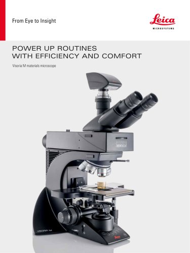
 Visoria M flyer
2 Pages
 Visoria M brochure
12 Pages
 Mateo FL Flyer EN
2 Pages
 DVM6 Brochure en
16 Pages
 Leica M50, M60, M80
12 Pages
 XL Stand
4 Pages
 LED1000
16 Pages
 LED3000 BLI
20 Pages
 LED5000 NVI
20 Pages
 LED3000 NVI
20 Pages
 LED3000 DI
20 Pages
 LED5000 HDI
20 Pages
 LED5000 CXI
20 Pages
 LED2500
8 Pages
 LED5000 MCI
20 Pages
 LED3000 MCI
20 Pages
 LED5000 SLI
20 Pages
 LED3000 SLI
20 Pages
 LED2000
8 Pages
 LED5000 RL
20 Pages
 LED3000 RL
20 Pages
 EL6000
4 Pages
 M320 F12 for ENT
12 Pages
 M620 TTS
4 Pages
 M220 F12
8 Pages
 RUV800
4 Pages
 DI C800
2 Pages
 M620 F20
8 Pages
 M822 F40 / F20
20 Pages
 M844 F40 / F20
16 Pages
 Proveo 8
16 Pages
 Envisu
8 Pages
 EnFocus
8 Pages
 M525 F20
12 Pages
 FL400
FL4004 Pages
 Leica M720 OH5
24 Pages
 Leica M530 OH6
16 Pages
 Leica M530 OHX
16 Pages
 Leica CaptiView
4 Pages
 Leica GLOW800
4 Pages
 Leica ARveo
16 Pages
 Leica PROvido
8 Pages
 LAS X Steel Expert
4 Pages
 Leica LMD Software
16 Pages
 Leica Map
12 Pages
 Leica Cleanliness Expert
4 Pages
 Leica LAS X Industry
4 Pages
 Leica LAS X Life Science
4 Pages
 Leica EM CPD300
12 Pages
 Leica EM TP
4 Pages
 Leica EM VCT500
2 Pages
 Leica EM ACE900
12 Pages
 Leica EM ACE200
12 Pages
 Leica EM KMR3
8 Pages
 Leica EM AFS2
8 Pages
 Leica EM ICE
12 Pages
 TCS SP8 DLS
16 Pages
 Leica LIGHTNING
2 Pages
 Leica SP8 DIVE
20 Pages
 Leica K5
2 Pages
 Leica DFC295
6 Pages
 Leica DFC3000 G
6 Pages
 Leica EC4
4 Pages
 Leica DFC7000 T / DFC7000 GT
4 Pages
 Leica DFC9000
2 Pages
 Leica MZ10 F
4 Pages
 Leica M165 FC
16 Pages
 Leica DM IL LED
12 Pages
 Leica DMi1
2 Pages
 THUNDER Imager Tissue
2 Pages
 Leica FS M
12 Pages
 Leica FS C
20 Pages
 Leica FS CB
20 Pages
 Leica DM3000 / DM3000 LED
16 Pages
 Leica DM750
12 Pages
 Leica DM500
12 Pages
 Leica DM300
8 Pages
 Leica DM1750 M
12 Pages
 Leica FS4000 LED
20 Pages
 Leica DM2000 / DM2000 LED
16 Pages
 Leica DM1000
16 Pages
 Leica DM1000 LED
16 Pages
 Leica DM2500 & DM2500 LED
16 Pages
 Leica DM4 B & DM6 B
16 Pages
 Leica DMC2900
6 Pages
 Leica DMC6200
8 Pages
 Leica DMC5400
4 Pages
 Leica Z6 APO
16 Pages
 Leica Z16 APO
16 Pages
 Leica M205 FCA / M205 FA
16 Pages
 Leica DM12000 M
8 Pages
 Leica DM8000 M
8 Pages
 Leica DM4 P, DM2700 P, DM750 P
12 Pages
 Leica EM ACE600
12 Pages
 Leica EM RAPID
8 Pages
 Leica EM TRIM2
8 Pages
 Leica EM TIC 3X
16 Pages
 Leica EM TXP
10 Pages
 Leica DM2700 M
12 Pages
 DMi8 M / C / A
12 Pages
 Leica A60 S / Leica A60 F
16 Pages
 SOLUTIONS FOR MATERIALS SCIENCE
12 Pages
 Leica StereoZoom line
20 Pages
 TL-Bases
12 Pages
 TCS SP8
20 Pages
 TCS SP8 STED 3X
24 Pages
 TCS SP8 Objective
24 Pages
 AOBS
AOBS16 Pages
 Leica DM3 XL
7 Pages
 Leica DMi8 S
12 Pages
 Leica M50/M60/M80
12 Pages
 Leica DM750 M
12 Pages
 Leica DM4 M/ DM6 M
12 Pages
 Leica A60 S/A60 F
16 Pages


- Digital imager
- Visible imager
- CMOS camera module
- Industrial camera module
- Nora LED lighting
- Nora analysis software
- Positioning table
- Nora Windows software
- Full-color camera system
- Translation stage
- USB camera module
- Monochrome camera module
- 3D software solution
- Nora industrial software
- Nora optical microscope
- HD camera module
- LED hand lamp
- Quality software
- Nora laboratory microscope