- Company
- Products
- Catalogs
- News & Trends
- Exhibitions
EnFocus
1 /8Pages

EnFocus
1 /8Pages
Catalog excerpts

OCT system for ophthalmic surgery SEE WHAT YOU’VE BEEN MISSING EnFocus intrasurgical OCT
Open the catalog to page 1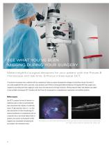
SEE WHAT YOU’VE BEEN MISSING DURING YOUR SURGERY Make insightful surgical decisions for your patient with the Proveo 8 microscope and real-time, EnFocus intrasurgical OCT. A top-down microscope view, combined with your experience, helps you assess intraoperative changes to subsurface tissues. But what if you could supplement this with a real-time, cross-sectional view? EnFocus intrasurgical Optical Coherence Tomograhy (OCT) can support your surgeries by providing real-time imaging of ocular tissue microstructures with high resolution, offering exquisite detail and deepest scan depth of any available...
Open the catalog to page 2
See even more: EnFocus with Proveo 8 Combine EnFocus OCT with the premium Proveo 8 microscope for a complete visualization platform for all your ophthalmic surgeries. The features and benefits of the Proveo 8 microscope include: Retina surgery > The BIOM 5 wide angle accessory from OCULUS with synchronized inversion and focusing optimizes fundus viewing > FusionOptics technology combines depth of field with increased resolution for a texture-rich view from the periphery to the retina Cornea surgery > usionOptics technology also aids in cornea surgery enabling F viewing of the complete anterior...
Open the catalog to page 3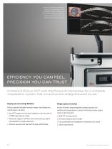
Large LCD for viewing of real-time OCT imaging, microscope image, and OCT scan position. Choose a simplified view for ease of use during your procedure or switch to full on-screen menu for comprehensive set-up and analysis tools. EFFICIENCY YOU CAN FEEL, PRECISION YOU CAN TRUST Combine EnFocus OCT with the Proveo 8 microscope for a complete visualization system that is intuitive and straightforward to use.* Display and record in High Definition Simply capture and analyze EnFocus captures the highest resolution images, then displays and records these in rich detail. Intuitive InVivoVue image management...
Open the catalog to page 4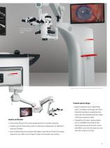
OCT image injection into binocular through the DIC800 Images courtesy of University Hospital Dusseldorf, Germany. Premium optical design > Optical innovations such as OptiChrome optics, FusionOptics technology and CoAx 4 illumination provide an outstanding microscope view with enhanced resolution, depth of field and constant red reflex Intuitive and flexible > Easily control EnFocus OCT and microscope functions via wireless footswitch > Combine with the Proveo 8 floor stand or ceiling mount configurations for flexibility to meet your OR needs > Smart workflow features and optical technologies...
Open the catalog to page 5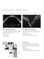
OUR QUALITY, YOUR CHOICE Innovative design with configurations to match your requirements. 11 mm Imaging Depth Image courtesy of Scott Oliver, MD, Director, Eye Cancer Program, University of Colorado EnFocus Ultra-Deep OCT: Deeper and wider EnFocus Ultra-HD OCT: Rich subsurface detail The EnFocus Ultra-Deep OCT option delivers very deep and wide imaging for full anterior segment visualization. Anterior structures continue to be resolved with crisp detail of ≤ 9 μm. EnFocus Ultra-HD OCT technology delivers extremely high definition, real-time images of either the posterior or the anterior segment....
Open the catalog to page 6
Microscope compatibility New or existing Proveo 8 microscopes, M844 microscopes Product Applications EnFocus Ultra-HD OCT High-Res Imaging Posterior, Anterior EnFocus Ultra-Deep OCT Deep Imaging Anterior, Posterior
Open the catalog to page 7
MC-0000531 · 18.02.2020 · EN · Copyright © 2019 Leica Microsystems (Schweiz) AG. All rights reserved. Subject to modifications. LEICA and the Leica Logo are registered trademarks of Leica Microsystems IR GmbH. XEN® is a registered trademark of AqueSys, Inc., an Allergan affiliate. EnFocus is a class IIa medical device 0123 Leica Microsystems NC, Inc. 4222 Emperor Blvd, Suite 390, Durham, NC 27703, USA Leica Microsystems (Schweiz) AG Max Schmidheiny-Strasse 201 9435 Heerbrugg, Switzerland Not all products or services are approved or offered in every market and approved labeling and instructions...
Open the catalog to page 8All Leica Microsystems GmbH catalogs and technical brochures
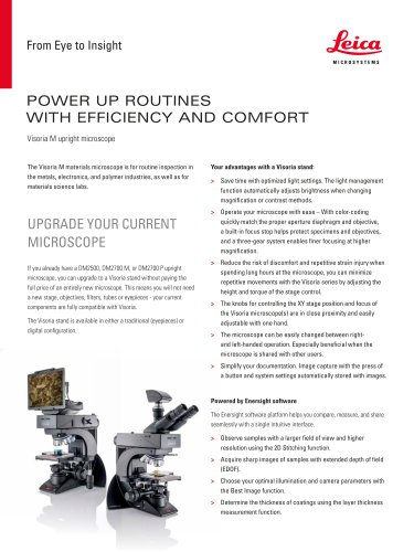 Visoria M flyer
2 Pages
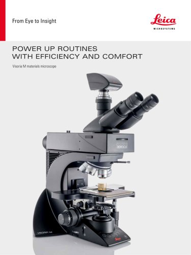 Visoria M brochure
12 Pages
 Mateo FL Flyer EN
2 Pages
 DVM6 Brochure en
16 Pages
 Leica M50, M60, M80
12 Pages
 XL Stand
4 Pages
 LED1000
16 Pages
 LED3000 BLI
20 Pages
 LED5000 NVI
20 Pages
 LED3000 NVI
20 Pages
 LED3000 DI
20 Pages
 LED5000 HDI
20 Pages
 LED5000 CXI
20 Pages
 LED2500
8 Pages
 LED5000 MCI
20 Pages
 LED3000 MCI
20 Pages
 LED5000 SLI
20 Pages
 LED3000 SLI
20 Pages
 LED2000
8 Pages
 LED5000 RL
20 Pages
 LED3000 RL
20 Pages
 EL6000
4 Pages
 M320 F12 for ENT
12 Pages
 M620 TTS
4 Pages
 M220 F12
8 Pages
 RUV800
4 Pages
 DI C800
2 Pages
 M620 F20
8 Pages
 M822 F40 / F20
20 Pages
 M844 F40 / F20
16 Pages
 Proveo 8
16 Pages
 Envisu
8 Pages
 M525 F20
12 Pages
 FL400
FL4004 Pages
 Leica M525 OH4
20 Pages
 Leica M720 OH5
24 Pages
 Leica M530 OH6
16 Pages
 Leica M530 OHX
16 Pages
 Leica CaptiView
4 Pages
 Leica GLOW800
4 Pages
 Leica ARveo
16 Pages
 Leica PROvido
8 Pages
 LAS X Steel Expert
4 Pages
 Leica LMD Software
16 Pages
 Leica Map
12 Pages
 Leica Cleanliness Expert
4 Pages
 Leica LAS X Industry
4 Pages
 Leica LAS X Life Science
4 Pages
 Leica EM CPD300
12 Pages
 Leica EM TP
4 Pages
 Leica EM VCT500
2 Pages
 Leica EM ACE900
12 Pages
 Leica EM ACE200
12 Pages
 Leica EM KMR3
8 Pages
 Leica EM AFS2
8 Pages
 Leica EM ICE
12 Pages
 TCS SP8 DLS
16 Pages
 Leica LIGHTNING
2 Pages
 Leica SP8 DIVE
20 Pages
 Leica K5
2 Pages
 Leica DFC295
6 Pages
 Leica DFC3000 G
6 Pages
 Leica EC4
4 Pages
 Leica DFC7000 T / DFC7000 GT
4 Pages
 Leica DFC9000
2 Pages
 Leica MZ10 F
4 Pages
 Leica M165 FC
16 Pages
 Leica DM IL LED
12 Pages
 Leica DMi1
2 Pages
 THUNDER Imager Tissue
2 Pages
 Leica FS M
12 Pages
 Leica FS C
20 Pages
 Leica FS CB
20 Pages
 Leica DM3000 / DM3000 LED
16 Pages
 Leica DM750
12 Pages
 Leica DM500
12 Pages
 Leica DM300
8 Pages
 Leica DM1750 M
12 Pages
 Leica FS4000 LED
20 Pages
 Leica DM2000 / DM2000 LED
16 Pages
 Leica DM1000
16 Pages
 Leica DM1000 LED
16 Pages
 Leica DM2500 & DM2500 LED
16 Pages
 Leica DM4 B & DM6 B
16 Pages
 Leica DMC2900
6 Pages
 Leica DMC6200
8 Pages
 Leica DMC5400
4 Pages
 Leica Z6 APO
16 Pages
 Leica Z16 APO
16 Pages
 Leica M205 FCA / M205 FA
16 Pages
 Leica DM12000 M
8 Pages
 Leica DM8000 M
8 Pages
 Leica DM4 P, DM2700 P, DM750 P
12 Pages
 Leica EM ACE600
12 Pages
 Leica EM RAPID
8 Pages
 Leica EM TRIM2
8 Pages
 Leica EM TIC 3X
16 Pages
 Leica EM TXP
10 Pages
 Leica DM2700 M
12 Pages
 DMi8 M / C / A
12 Pages
 Leica A60 S / Leica A60 F
16 Pages
 SOLUTIONS FOR MATERIALS SCIENCE
12 Pages
 Leica StereoZoom line
20 Pages
 TL-Bases
12 Pages
 TCS SP8
20 Pages
 TCS SP8 STED 3X
24 Pages
 TCS SP8 Objective
24 Pages
 AOBS
AOBS16 Pages
 Leica DM3 XL
7 Pages
 Leica DMi8 S
12 Pages
 Leica M50/M60/M80
12 Pages
 Leica DM750 M
12 Pages
 Leica DM4 M/ DM6 M
12 Pages
 Leica A60 S/A60 F
16 Pages


- Digital imager
- Visible imager
- CMOS camera module
- Industrial camera module
- Nora LED lighting
- Nora analysis software
- Positioning table
- Full-color camera system
- Nora Windows software
- Translation stage
- USB camera module
- Monochrome camera module
- 3D software solution
- Nora industrial software
- Nora optical microscope
- HD camera module
- LED hand lamp
- Quality software
- Nora laboratory microscope