JSM-F100
1 /20Pages

JSM-F100
1 /20Pages
Catalog excerpts

Scientific / Metrology Instruments Schottky Field Emission Scanning Electron Microscope The next level of analytical intelligence in FE-SEM
Open the catalog to page 1
Introducing FE-SEM: Integrating Optical Image, SEM Image, and EDS Analysis JSM-F100 The JSM-F100 navigates you to the world of unknown nanostructures.
Open the catalog to page 2
Optical image Intensity [Counts] Specimen: Electric lamp filament *Use of optical microscope requires an o
Open the catalog to page 3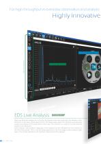
For high throughput in everyday observation and analysis Highly Innovative Zeromag (Optical Image) Spectrum Monitor Spectrum Monitor enables you to locate the analysis area with automatic live display of the elements in the area. While searching for the target area, Spectrum Monitor automatically makes live display of the elements in the area. This function is useful for screening the object to be measured. Point analysis, area analysis, mapping or line analysis can be selected from the buttons at the left of the screen. Then, click the target position on the live image to start analysis right...
Open the catalog to page 4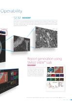
Zeromag is incorporated for seamless transition from optical to SEM image. This function is useful for locating the specimen area and for managing acquired data. The JSM-F100 provides not only high operability but also high-resolution images obtained with the FE-SEM. Report generation Report generation using SMILE VIEW Lab TM → P 12 The JSM-F100 offers connectivity with SMILE VIEW™ Lab, the new data management system used for JEOL's analytical instruments. It is useful for data management, analysis, and report generation. *The optional stage navigation system (SNS) is required for use of optical...
Open the catalog to page 5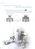
SEM A Variety of Functions for Multi-purpose Analysis In-lens Schottky Plus Field Emission electron Gun (FEG) The In-lens Schottky Plus FEG has realized improved brightness as a result of integration of the electron gun and low-aberration condenser lens. With this FEG, generated electrons can be efficiently focused, enabling probe currents on the order of a few pA to several tens of nA even at low accelerating voltages. High-resolution observation is easy: there is no need to switch the objective aperture for tasks from fast elemental mapping to EBSD to SXES (see P15) analyses. In-lens Schottky...
Open the catalog to page 6
The JSM-F100 comes with JEOL's electrostatic/Magnetic field superposed objective lens, Hybrid Lens (HL). This powerful lens enables observation and analysis of any specimen, including magnetic and insulating materials at high spatial resolution. Standard mode Magnetic lens effect Magnetic lens effect Magnetic lens effect Electrostatic lens effect Beam Deceleration (BD) mode Applying a bias voltage of up to −2 kV to the specimen stage enables deceleration of the incident electron beam just before the specimen. This function improves the spatial resolution and signal-to-noise (S/N) ratio at low...
Open the catalog to page 7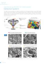
SEM Acquiring various information for multiple purposes Detector System The JSM-F100 accommodates four detectors, enabling you to simultaneously observe various SEM images uniquely formed by each detector. Like the general-purpose SEM, the JSM-F100 offers a Secondary Electron Detector (SED) and a Retractable Backscattered Electron Detector (RBED). In addition, this FE-SEM is configured with two Through The Lens (TTL) detectors: the Upper Electron Detector (UED) and the Upper Secondary Electron Detector (USD). * RBED and USD are optional 3D image obtained with SMILE VIEW Map *Option (Upper Electron...
Open the catalog to page 8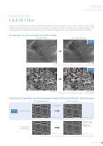
LIVE-AI Filter JEOL has incorporated the LIVE-AI(Artificial Intelligence)filter for higher quality of live images. Unlike image integration processing, the LIVE-AI filter can display a seamless moving live image with no residual image. This unique feature is very effective in searching for the observation area and for focus and astigmatism correction. Comparison of normal mode and LIVE-AI filter Normal mode With LIVE-AI filter AI filter 1 μm 1 μm Specimen: Ant exoskeleton, Accelerating voltage: 0.5 kV, Detector: SED 0.5 μm Specimen: Iron rust, Accelerating voltage: 1 kV, Detector: SED Comparison...
Open the catalog to page 9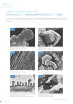
SEM Capable of nanostructure observation Examples of High-spatial-resolution Images The JSM-F100 FE-SEM permits observation of nanostructures. Selecting observation conditions and detectors suitable for your applications enables you to acquire characteristic SEM images from a variety of specimens. Specimen: Pt nanoparticles on carbon, Accelerating voltage: 20 kV, WD: 2 mm, Observation mode: BD, Detector: UED Specimen: Seal tape, Accelerating voltage: 0.5 kV, WD: 2 mm, Observation mode: BD, Detector: UED Specimen: Zeolite, Accelerating voltage: 1 kV, WD: 3 mm, Observation mode: STD, Detector:...
Open the catalog to page 10
EDS Immediate elemental analysis of observation areas EDS Integration With next-generation operability realized, you can transition seamlessly from SEM imaging to elemental analysis by EDS. Preselect area, mapping, line, or another type of analysis directly on the observation screen to begin analyzing your specimen immediately. Observation screen Check the analysis position using Live Analysis. Specify analysis ② position. Select analysis *Preselect analysis positions on multiple areas: each analysis will begin automatically, enabling you to obtain multiple measurements. Specimen: Cross section...
Open the catalog to page 11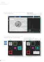
Elemental screening for EDS analysis always available Live Analysis Spectrum Monitor : Always displays a spectrum of the constituent elements in the observation area. Live image Specimen: Cross section of cast iron. For reducing errors in analysis during acquisition You can switch between screens during map acquisition. Eliminates background effects Effects of peak Mn and Fe overlaps are avoided. Net count map: Reflects analysis results with automatic background removal and peak deconvoluted maps. Useful when the spectrum contains peak overlaps or very small peaks.
Open the catalog to page 12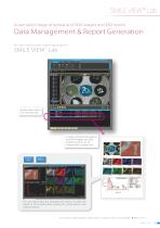
Automatic linkage of optical and SEM images and EDS results Data Management & Report Generation For fast and simple report generation Simply select items from the data list. A report compiling the optical and SEM images and EDS analysis results can be created with a single click. You can easily edit and reanalyze the report: its data are linked to the original data acquisition, observation, and analysis results. *The optional stage navigation system (SNS) is required for use of optical images. ➡ Refer to P.16.
Open the catalog to page 13All Jeol catalogs and technical brochures
 Products Guide 2023
24 Pages
 CROSS SECTION POLISHER
4 Pages
 Super Spectrometer
4 Pages
 JSM-7610Plus
4 Pages
 JEM-ARM300F2
2 Pages
 JCM 6000 Catalogue
16 Pages
 SpiralTOF JMS-S3000
16 Pages

Archived catalogs
 JST-F Series Power Supplies
16 Pages


- AMOT tank
- Automation software solution
- Concentration analyzer
- AMOT analysis software
- AMOT process software
- AMOT Windows software
- AMOT automatic analyzer
- AMOT spectrometer
- AMOT feeder
- Process analyzer
- AMOT 3D software
- AMOT industrial software
- AMOT interface software
- Industrial tank container
- Additive manufacturing machine
- AMOT visualization software
- Real-time analyzer
- AMOT laboratory analyzer
- AMOT laboratory microscope