- Catalogs
- Imagine Optic
- Simple method to improve image contrast in spinning disk microscopy using adaptive optics - Adaptive optics for microscopy Application Notes
Simple method to improve image contrast in spinning disk microscopy using adaptive optics - Adaptive optics for microscopy Application Notes
1 /4Pages
Simple method to improve image contrast in spinning disk microscopy using adaptive optics - Adaptive optics for microscopy Application Notes
1 /4Pages
Catalog excerpts
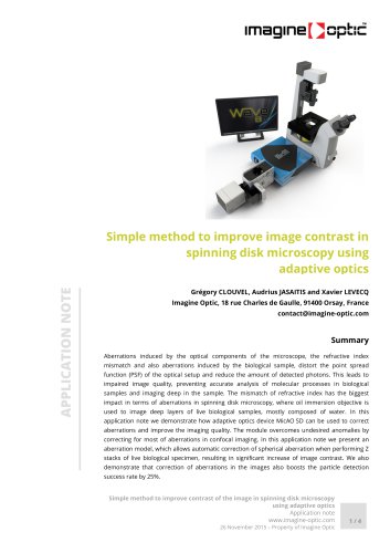
Simple method to improve image contrast in spinning disk microscopy using adaptive optics (Choose the color according to product type) Grégory CLOUVEL, Audrius JASAITIS and Xavier LEVECQ Imagine Optic, 18 rue Charles de Gaulle, 91400 Orsay, France [email protected] Summary Aberrations induced by the optical components of the microscope, the refractive index mismatch and also aberrations induced by the biological sample, distort the point spread function (PSF) of the optical setup and reduce the amount of detected photons. This leads to impaired image quality, preventing accurate analysis of molecular processes in biological samples and imaging deep in the sample. The mismatch of refractive index has the biggest impact in terms of aberrations in spinning disk microscopy, where oil immersion objective is used to image deep layers of live biological samples, mostly composed of water. In this application note we demonstrate how adaptive optics device MicAO SD can be used to correct aberrations and improve the imaging quality. The module overcomes undesired anomalies by correcting for most of aberrations in confocal imaging. In this application note we present an aberration model, which allows automatic correction of spherical aberration when performing Z stacks of live biological specimen, resulting in significant increase of image contrast. We also demonstrate that correction of aberrations in the images also boosts the particle detection success rate by 25%. Simple method to improve contrast of the image in spinning disk microscopy using adaptive optics Application note www.imagine-optic.com 26 November 2015 – Property of Imagine Optic
Open the catalog to page 1
Spinning disk (SD) microscopy is one of the most common imaging techniques with optical sectioning capability used in research laboratories for live imaging. This technology represents a state-of-theart approach to study morphogenesis. It can resolve live biological processes on a multitude of spatial scales from sub-cellular to relatively thick tissues. SD induces low photo damage and allows for fast multicolor 3D imaging using a low-noise and highdynamic-range detector such as EMCCD or sCMOS camera. Nevertheless, this type of microscopy is affected by aberrations, induced by the optical elements...
Open the catalog to page 2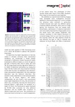
Figure 2. Comparison of gamma-tubulin-GFP on fixed Drosophila imaginal disk (centrosomes of epithelial cells) acquired with spinning disk microscope with (+AO) or without MicAO SD (-AO). A. Images on the left are the maximum intensity projections of 25 µm Z stack in XY plane. Images on the right show the XZ view of the stack. B. Comparison of wavefront measurements with and without AO at different depths. At each depth, the profile line with normalized intensities reveals improvement achieved with AO. The scale bar is for both XY and YZ. model has been applied in STED microscopy (Lenz et al.,...
Open the catalog to page 3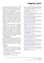
correction was even better, resulting in a 2-fold improvement. Moreover, at depth the shape of the centrosome is also corrected and better preserved, allowing for a much better assessment and interpretation of the observed sample. Similar results we also obtained by imaging live Drosophila brain samples (Figure 3) – the contrast of the images is significantly improved. In this case we decided to verify if the correction of aberrations can improve the statistics of particle detection. For the detection of centrosomes we used Spot Detector tool from Icy imaging software package (Institut Pasteur,...
Open the catalog to page 4All Imagine Optic catalogs and technical brochures
 WAVE Suite
3 Pages


Archived catalogs
 HASO™3 Wavefront Sensors
3 Pages
 SL-Sys neo
2 Pages
 SL-Sys LIQUID
2 Pages
 HASO R-Flex
3 Pages
 HASO?3 WSR Wavefront Sensors
2 Pages
 bendAO?
3 Pages
 HASO3
HASO32 Pages
 HASO R.FLEX
4 Pages
 absolute measurement
4 Pages
 Telescope characterization
3 Pages
 NIR optics characterization
6 Pages
 Large deformable mirror ILAO
6 Pages
 AO in femtosecond laser
5 Pages
 AO inside laser chain
5 Pages
 Microtraps
4 Pages