Quasi-isotropic nanometric 3D resolution in PALM/STORM with the help of MicAO 3DSR - Adaptive optics for microscopy Application Notes
1 /
6Pages
Catalog excerpts

Quasi-isotropic nanometric 3D resolution in PALM/STORM with the help of MicAO 3DSR Grégory CLOUVEL, Audrius JASAITIS and Xavier LEVECQ Imagine Optic, 18 rue Charles de Gaulle, 91400 Orsay, France contact@imagine-optic.com Summary Photoactivation localization microscopy (PALM) and stochastic optical reconstruction microscopy (STORM) are becoming routine methods in biological imaging with optical resolution beyond the light diffraction barrier. We present MicAO 3DSR – the first adaptive optics device which introduces the three dimensional imaging capability for PALM/STORM. This device contains a wavefront sensor and a deformable mirror that enables it to correct various types of aberrations including those induced by the optical elements inside the microscope and by the biological sample itself. In this way, MicAO optimizes the Point Spread Function (PSF) of the microscope and consequently improves the lateral localization precision of the PALM/STORM method. In addition to that, MicAO 3D-SR provides the ability to image fluorescent molecules in 3D. To achieve this, it uses its deformable mirror to introduce controlled perfect astigmatism and thereby allows us to precisely locate the position of fluorescing molecule in all three dimensions. In this application note we demonstrate that by optimizing the PSF with MicAO 3DSR and after introduction of perfect astigmatism, we are able to reach almost isotropic nanometric resolution in PALM/STORM method. For example, at 1000 detected photons, the typical photon flux in PALM/STORM method, we demonstrate quasi-isotropic localization precision of 8 nm in lateral and 16 nm in axial directions. Quasi-isotropic nanometric 3D resolution in PALM/STORM with the help of MicAO 3DSR Application note www.imagine-optic.com 8 December 2015 – Property of Imagine Optic
Open the catalog to page 1
PALM and STORM are becoming more and more popular techniques in the field of super resolution fluorescence microscopy (Betzig et al, 2006; Hess et al, 2006; Rust et al, 2006). These single-molecule localization methods are easy to implement on standard microscopes and they provide the best resolution among other superresolution methods, such as structured illumination microscopy (SIM, Gustafsson, 2005) or stimulated emission depletion microscopy (STED, Hell and Wichmann, 1994). In PALM/STORM methods, the assumption that the detected fluorescence is originating from a single fluorophore...
Open the catalog to page 2
Dissipation of useful photons by aberrations is demonstrated in Figure 1 with the use of numerical simulations of spherical aberration. In this example we introduced a spherical aberration with the amplitude of 67 nm root mean square (RMS) – the common type of aberration occurring when animal tissue culture cells are imaged with a microscope (Schwertner et al, 2007). In this case the spherical aberration is caused by the mismatch of refractive indexes between water in the biological sample and the immersion oil of the objective. Even though the total number of photons in both images is the...
Open the catalog to page 3
Figure 5. The calibration curve that is used for the determination of the location of the fluorophore along the z axis. compare the intensity profiles. The maximum value of the intensity profile is increased by 90% after optimizations. As it will be discussed later, this will significantly improve the resolution of the PALM/STORM setup. To correct for the aberrations induced by the biological sample, we introduced a small drop (20-50 µl) of 100 nm diameter multicolor fluorescent beads -5 -4 (Invitrogen, 10 -10 dilution) directly into the sample. We then washed the sample twice with the...
Open the catalog to page 4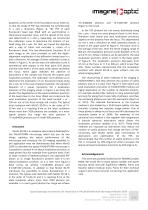
positions: at the center of the focal plane and at ±250 nm, so that the shape of PSF was extended not symmetrically in x and y directions (Figure 4). The PSF of each fluorescent bead was fitted with an asymmetrical 2dimensional Gaussian curve, and the spread of the curve was determined in x and y. Separately, we constructed the calibration curve to determine the location of our bead along the z axis. For that, we moved the objective with a step of 24nm and recorded a z-stack of a fluorescent bead. The two-dimensional Gaussian fit of each image in the stack provided us with the...
Open the catalog to page 5
References Abrahamsson S, Chen J, Hajj B, Stallinga S, Katsov AY, Wisniewski J, Mizuguchi G, Soule P, Mueller F, Darzacq CD, Darzacq X, Wu C, Bargmann CI, Agard DA, Dahan M, Gustafsson MGL (2012) Fast multicolor 3D imaging using aberration-corrected multifocus microscopy Nat. Met. 10, 60-63. Betzig E, Patterson GH, Sourgat R, Lindwaser OW, Olenych S, Bonifacino JS, Davidson MW, LippincottSchwartz J, Hess HF (2006) Imaging intracellular fluorescent proteins at nanometer resolution. Science, 313, 1642-1645. Booth MJ, Neil MAA Juskaitis R and Wilson T (2002) Adaptive aberration...
Open the catalog to page 6All Imagine Optic catalogs and technical brochures
-
WAVE Suite
3 Pages
Archived catalogs
-
Microtraps
4 Pages
-
AO inside laser chain
5 Pages
-
AO in femtosecond laser
5 Pages
-
Large deformable mirror ILAO
6 Pages
-
NIR optics characterization
6 Pages
-
Telescope characterization
3 Pages
-
absolute measurement
4 Pages
-
HASO R.FLEX
4 Pages
-
HASO3
2 Pages
-
bendAO?
3 Pages
-
HASO?3 WSR Wavefront Sensors
2 Pages
-
HASO R-Flex
3 Pages
-
SL-Sys LIQUID
2 Pages
-
SL-Sys neo
2 Pages
-
HASO™3 Wavefront Sensors
3 Pages

