- Catalogs
- Imagine Optic
- Deep 3D PALM/STORM imaging: MicAO 3DSR – the key to combining depth and highest resolution - Adaptive optics for microscopy Application Notes
Deep 3D PALM/STORM imaging: MicAO 3DSR – the key to combining depth and highest resolution - Adaptive optics for microscopy Application Notes
1 /5Pages
Deep 3D PALM/STORM imaging: MicAO 3DSR – the key to combining depth and highest resolution - Adaptive optics for microscopy Application Notes
1 /5Pages
Catalog excerpts

Deep 3D PALM/STORM imaging: MicAO 3DSR – the key to combining depth and highest resolution Grégory CLOUVEL, Audrius JASAITIS and Xavier LEVECQ Imagine Optic, 18 rue Charles de Gaulle, 91400 Orsay, France [email protected] Summary The determination of 3D arrangement of cellular structures has become a necessary requirement in cellular biology. Unfortunately, the size of such structures usually lies beyond the diffraction limit and therefore they cannot be visualized in studies using today’s widely popular fluorescence microscopy techniques. Photoactivation localization microscopy (PALM) and stochastic optical reconstruction microscopy (STORM) enables us to locate fluorescent molecules with nanometric resolution. Unfortunately in current implementations, these techniques are efficient only in the vicinity of the coverslip, like in total internal reflection (TIRF) or in the first few micrometers inside the sample. Imaging deeper is highly perturbed by the spherical aberration, which is caused by the refractive index mismatch between the sample and immersion oil of the objective. Aberrations can be efficiently corrected using adaptive optics. Here for the correction of aberrations we used MicAO 3DSR – an adaptive optics device containing Shack-Hartman-type wavefront sensor and continuous membrane deformable mirror. By correcting the spherical aberration we can obtain perfectly symmetrical PSF along the Z axis at the depth reaching 50µm in the sample. After the aberration correction, MicAO 3DSR can apply variable amount of astigmatism for three-dimensional imaging. To test the performance of this system for deep PALM imaging we constructed a model sample composed of fixed HeLa cells, stably expressing centrosomal protein centrin-1. For optimization and drift correction, we also added to the sample 100nm fluorescent beads. Our results show that, after correction of aberrations, single molecule imaging can be performed at depths reaching 50µm. Deep 3D PALM/STORM imaging with MicAO 3DSR Application note www.imagine-optic.com 8 December 2015 – Property of Imagine Op
Open the catalog to page 1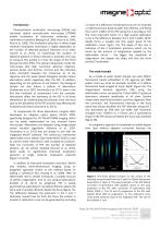
Introduction Photoactivation localization microscopy (PALM) and stochastic optical reconstruction microscopy (STORM) enable localization of fluorescent molecules with nanometric resolution (Betzig et al, 2006; Hess et al, 2006; Rust et al, 2006). The localization precision in these single molecule localization techniques is highly dependent on the number of detected photons (Thomson et al, 2002; Clouvel at al, 2013), so maximizing the number of detected photons in these methods is essential. One way to measure this quality is in fact the shape of the Point Spread Function (PSF). The optical components...
Open the catalog to page 2
extensions of the PSF (the upper panel of figure 3B). However, correction of aberrations completely restores the axial symmetry. As seen in figure 3C, when we corrected for 165nm RMS of spherical aberration and applied 90nm RMS of astigmatism, the PSF became laterally extended as needed above and below the focus, and the resulting calibration curve is perfectly symmetrical. Figure 2. The Z profile of the 200nm fluorescent bead at 22µm depth in 2% agarose. A. Before and after correction of aberrations. The size of the scalebar is 1µm; B. The intensity profile along the Z axis before (blue) and...
Open the catalog to page 3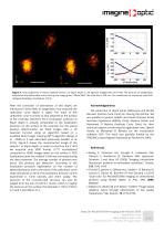
Figure 4. Two projections of mEos2 labeled Centrin1 at 50µm depth in 2% agarose imaged with 3D PALM. The amount of astigmatism induced on the deformable mirror during the imaging was 130nm RMS. The scale bar is 200 nm. For visualization of results we used ViSP software (El Beheiry and Dahan, 2013). After the correction of aberrations at this depth, we introduced 130nm RMS of astigmatism and acquired the calibration curve (figure 4, right). The slope of this calibration curve is similar to that obtained at the surface of the coverslip, therefore the Z localization precision at 50µm depth is actually...
Open the catalog to page 4
El Beheiry M and Dahan M (2013) “ViSP: representing single-particle localizations in three dimensions,” Nat. Meth., 10, 689-690. Facomprez A, Beaurepaire E and Débarre D (2012) “Accuracy of correction in modal sensorless adaptive optics,” Opt. Express, 20, 2598-2612. Hess ST, Grirajan TPK and Mason MD (2006) “Ultra-high resolutionimaging by fluorescence photoactivation localization microscopy,” Biophys. J., 91, 4258-4272. Huang B, Wang J, Bates M and Zhuang X (2008) “Threedimensional super-resolution imaging by stochastic optical reconstruction microscopy,” Science, 319, 810813. Izeddin...
Open the catalog to page 5All Imagine Optic catalogs and technical brochures
 WAVE Suite
3 Pages


Archived catalogs
 HASO™3 Wavefront Sensors
3 Pages
 SL-Sys neo
2 Pages
 SL-Sys LIQUID
2 Pages
 HASO R-Flex
3 Pages
 HASO?3 WSR Wavefront Sensors
2 Pages
 bendAO?
3 Pages
 HASO3
HASO32 Pages
 HASO R.FLEX
4 Pages
 absolute measurement
4 Pages
 Telescope characterization
3 Pages
 NIR optics characterization
6 Pages
 Large deformable mirror ILAO
6 Pages
 AO in femtosecond laser
5 Pages
 AO inside laser chain
5 Pages
 Microtraps
4 Pages