- Catalogs
- Imagine Optic
- 3D dual-color PALM/dSTORM imaging of centrosomal proteins with nanometric resolution using MicAO 3DSR - Adaptive optics for microscopy Application Notes

3D dual-color PALM/dSTORM imaging of centrosomal proteins with nanometric resolution using MicAO 3DSR - Adaptive optics for microscopy Application Notes

3D dual-color PALM/dSTORM imaging of centrosomal proteins with nanometric resolution using MicAO 3DSR - Adaptive optics for microscopy Application Notes
Photoactivation localization microscopy (PALM) and stochastic optical reconstruction microscopy (STORM) are advanced imaging techniques that overcome the light diffraction barrier, enabling high-resolution biological imaging. The MicAO 3DSR device, developed by Imagine Optic, enhances these methods by introducing three-dimensional imaging capabilities and correcting optical aberrations, improving localization precision by 40%.
Fluorescence microscopy is essential for visualizing intracellular components, but traditional methods are limited by the diffraction barrier. Super-resolution techniques like PALM and STORM have emerged to overcome this, achieving resolutions as fine as 10 nm. However, these methods traditionally lack precise axial information for 3D imaging.
The MicAO 3DSR device uses adaptive optics, including a wavefront sensor and deformable mirror, to correct aberrations and optimize the Point Spread Function (PSF). This results in improved photon detection and localization precision. The device can introduce controlled astigmatism, enabling 3D imaging similar to cylindrical lenses but without additional aberrations.
The PALM/STORM setup was based on a Nikon Ti-E microscope with MicAO 3DSR inserted between the microscope and camera. Initial optimization involved aligning the device using fluorescent beads and correcting aberrations with a closed-loop system. Biological samples were further optimized for aberrations using a 3N algorithm.
Using MicAO 3DSR, the localization precision of PALM/STORM was improved by 40%, with lateral resolutions of 5-8 nm and axial resolutions of 10-16 nm. The device enabled 3D dual-color imaging of centrosomal proteins, achieving a localization precision of 7 nm laterally and 13 nm axially.
MicAO 3DSR significantly enhances the capabilities of PALM/STORM imaging by providing high-resolution 3D imaging of cellular structures, offering a promising alternative to traditional methods like immuno-electron microscopy.
- Centrin staining in the mother-centriole appears tilted by approximately 40 degrees relative to the distal appendage ring.
- A top view of the mother-centriole shows an elongated centrin concentration, suggesting a complex spatial distribution.
- The Cep164 ring was incomplete, possibly due to technical reasons, and appeared to consist of straight elements potentially corresponding to distal appendages.
The study utilized PALM/STORM setup in conjunction with MicAO 3D-SR to achieve high-resolution imaging of centrosomal proteins.
The preliminary results demonstrate the effectiveness of the imaging setup, though further research with additional centrosomal markers is necessary.
The research was partially funded by the TRIDIMIC project and involved contributions from various researchers and institutions.
The document cites numerous studies related to super-resolution microscopy and centrosomal protein localization, providing a comprehensive background for the research.
Catalog excerpts

3D dual-color PALM/dSTORM imaging of centrosomal proteins with nanometric resolution using MicAO 3DSR Grégory CLOUVEL, Audrius JASAITIS and Xavier LEVECQ Imagine Optic, 18 rue Charles de Gaulle, 91400 Orsay, France [email protected] Summary Photoactivation localization microscopy (PALM) and stochastic optical reconstruction microscopy (STORM) are becoming routine methods in biological imaging with optical resolution beyond the light diffraction barrier. We present MicAO 3DSR – the first adaptive optics device which introduces the three dimensional imaging capability for PALM/STORM. This apparatus, designed by Imagine Optic, contains the wave-front sensor and deformable mirror. With the help of these components MicAO 3D-SR corrects various types of aberrations, induced by optical elements inside the microscope and by the biological sample. MicAO 3D-SR optimizes the Point Spread Function (PSF) of the microscope and consequently improves the lateral localization precision of the PALM/STORM setup by 40%. In addition to that, MicAO 3D-SR brings in the ability to image fluorescent molecules in 3D. With its deformable mirror, it introduces controlled perfect astigmatism and allows us to precisely locate the position of fluorescing molecule in all three dimensions. In this application note we demonstrate the localization precision of PALM/STORM method when it is operated together with MicAO 3D-SR by 3D dual-color imaging of two proteins in centrosome – the cellular organelle which dimensions lye beyond the diffraction limit of 250x250x500 nm. 3D dual-color PALM/dSTORM imaging of centrosomal proteins with nanometric resolution using MicAO 3DSR Application note imagine-optic.com 8 December 2015 – Property of Imagine Optic
Open the catalog to page 1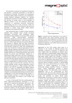
The nanometric resolution of intracellular components has become a necessary requirement in the field of fluorescence microscopy. The question of co-localization of proteins within cellular structures is very important in solving important biological problems. For example visualizing the structure of various adhesion complexes, the structure of nuclear pores, or the localization of different proteins in the cell membrane would help researchers to understand functional mechanisms. Unfortunately, the size of such structures usually lies beyond the diffraction limit and therefore they cannot be...
Open the catalog to page 2
Figure 2. Image (averaged over 40 individual beads) of the PSF with 60 nm RMS astigmatism at -250 nm of the focus (left image), at the focus (center image), and at +250 nm of the focus (right image). Centrosome plays an important role in many different cellular processes including the organization of microtubule arrays, cell cycle progression, the establishment of polarity of the cell and formation of primary cilium. One centrosome consists of two centrioles, which are barrel-shaped structures made of microtubules that are approximately 200 nm in diameter and 400–500 nm in length (Robbins et...
Open the catalog to page 3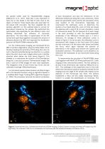
the specific buffer used for PALM/dSTORM imaging (Sillibourne et al , 2011). Note that it was important to have one or two beads in the field of view close to the region of interest. These beads were also used later for the lateral drift correction. We then reapplied the 3N iterative algorithm on the selected bead to correct for aberrations induced by the sample. We performed this optimization step separately for two different colors and thereby eliminated the influence of chromatic aberrations. The total effect of optimization of the PSF (closed and open-loop together) increased the amount of...
Open the catalog to page 4
Cep164 component of distal appendages was also chosen as a candidate for simultaneous dSTORM imaging because distal appendages of the mother centrioles are only a few nanometers in width and are challenging to image by immuno-EM. The HeLa mEos2-centrin1 cell line was labeled with an antibody to Cep164, stained with Alexa 647-conjugated secondary antibody and dual color two dimensional PALM/dSTORM imaging was performed (Sillibourne et al, 2011). In this application note, we present the dual-color 3-dimensional imaging of the same centriolar proteins using MicAO 3D-SR. Two different orientations...
Open the catalog to page 5
Sillibourne JE, Specht CG, Izeddin I, Hurbain I, Tran P, Triller A, Darzacq X, Dahan M, Bornens M. (2011) Assessing the Localization of Centrosomal Proteins by PALM/STORM Nanoscopy. Cytoskeleton 68, 619-627. Vorobjev IA, Chentsov Yu S. (1982) Centrioles in the cell cycle. I. Epithelial cells. J Cell Biol, 93, 938–949. Quirin S, Pavani SRP, Piestun R. (2012) Optimal 3D singlemolecule localization for super resolution microscopy with aberrations and engineered point spread functions. PNAS, 109, 675-679. 3D dual-color PALM/dSTORM imaging of centrosomal proteins with nanometric resolutionusing...
Open the catalog to page 6All Imagine Optic catalogs and technical brochures
 WAVE Suite
3 Pages







Archived catalogs
 HASO™3 Wavefront Sensors
3 Pages
 SL-Sys neo
2 Pages
 SL-Sys LIQUID
2 Pages
 HASO R-Flex
3 Pages
 HASO?3 WSR Wavefront Sensors
2 Pages
 bendAO?
3 Pages
 HASO3
HASO32 Pages
 HASO R.FLEX
4 Pages
 absolute measurement
4 Pages
 Telescope characterization
3 Pages
 NIR optics characterization
6 Pages
 Large deformable mirror ILAO
6 Pages
 AO in femtosecond laser
5 Pages
 AO inside laser chain
5 Pages
 Microtraps
4 Pages