- Catalogs
- HORIBA Scientific
- iHR550 Imaging Spectrometer
iHR550 Imaging Spectrometer
1 /4Pages

iHR550 Imaging Spectrometer
1 /4Pages
Catalog excerpts

Imaging Spectrometer OSD-IS-01 Simply the Best Imaging Spectrometer with No Compromise The iHR550 imaging spectrometer from HORIBA Scientific is simply the most versatile spectrometer on the market with no compromise among imaging, spectroscopy, and adaptability. The iHR550 utilizes a unique patented asymmetric design, which provides superior image quality and minimizes unwanted optical aberrations common to symmetric and crossed-Czerny Turner designs. For unrestricted flexibility, the iHR550 allows the user to take full advantage of the instrument by having two entrance and two exit ports for enhanced measurement capabilities. The iHR550 is the most suitable imaging spectrometer solution for: • Multi-track spectroscopy with tens of fibers imaged at once • Direct coupling for microscopy and 2D imaging • Hyperspectral imaging for Raman and luminescence applications • UV-Vis, near-IR and mid-IR spectroscopy with multiple array or single-channel detectors Virtually No Astigmatism When spatial resolution is needed, optical aberrations and—more precisely—astigmatism limits the imaging capabilities of the spectrometer. In a multi-track spectroscopy setup, where high spatial resolution is needed, astigmatism leads to a "bow-tie" effect, in which the image of each fiber blurs in the vertical direction towards the edge of the CCD. The iHR550 spectrometer minimizes astigmatism and delivers a sharp image of each fiber across the entire focal plane, as shown in Figure 1. Negligible Crosstalk To assess the degree of crosstalk between fibers, it is necessary to perform a horizontal bin of the full image. In a poorly designed spectrometer with a high degree of astigmatism, the signal between fibers begins to overlap, preventing clear separation between the fibers. The design of the iHR550 minimizes crosstalk between channels and improves contrast ratio. Figure 2 (next page) shows that the iHR550 image quality provides distinct peak separation. Figure 1. Image of a broadband quartz tungsten-halogen spectrum recorded with nineteen 200 μm fibers using the 1× imaging adapter with an iHR550 spectrometer, 1200 gr/mm grating blazed at 500 nm, and 1024×256 open-electrode CCD.
Open the catalog to page 1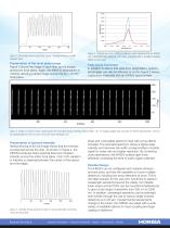
Figure 2: Horizontally binned results from Figure 1 showing minimal crosstalk between fibers. Figure 5: Spectral line from a mercury calibration lamp measured with an iHR550 and 1024×256 open-electrode CCD (blue), compared with a simulation showing effects of coma (red). Preservation of the focal plane image Figure 3 shows the image of each fiber as it is moved across the focal plane. Again, the effect of astigmatism is minimal, allowing a sharp image across the 30 × 12 mm focal plane. Fully Coma Corrected In addition to effects that arise from astigmatism, spectra and images can also be affected...
Open the catalog to page 2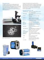
Microspectroscopy In addition to being the most suitable spectrometer for spectroscopy applications, the iHR550 imaging spectrometer offers outstanding performance when used for microspectroscopy. The iHR550 interfaces seamlessly with most commercially available microscopes as depicted in Figure 6 (top) on an inverted microscope for Raman spectroscopy. Imaging of the sample may be recorded with the microscope's witness camera or through the spectrometer itself, eliminating the need for an additional camera. Figure 6 (bottom) shows the image of Convallaria cells recorded with a grating tuned to...
Open the catalog to page 3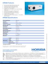
Up to four ports (two entrance and two exit) Kinematic turret with easy access hatch High-speed USB and additional hub port Purge port for UV and near-IR Easy CCD focus and alignment with external locking mechanism Choice of CCD or exit slit on either exit port Fast scanning capability: up to 160 nm/s Powerful SynerJY™ software for Windows® Optional internal filter wheel Choice of 2 mm slits for high resolution or 7 mm slits for high throughput iHR550 Specifications Focal length (mm) Grating size three, on-axis Flat field size Stray light Step size Length Width Optical axis Nominal weight Computer...
Open the catalog to page 4All HORIBA Scientific catalogs and technical brochures
 SLFA-60.6000
4 Pages
 PX-375
6 Pages
 XploRA Series Raman
8 Pages
 LabRAM Odyssey - Pre booking
2 Pages
 OMNI TERS Probes
2 Pages
 NanoRaman
7 Pages
 51 series
6 Pages
 LabRAM HR Evolution
5 Pages
 QPrep Automatic Dilutor
2 Pages
 KiloArc
2 Pages
 PowerArc
2 Pages
 Laqua
Laqua24 Pages
 Duetta
8 Pages
 MacroRAM
2 Pages
 PSA300
5 Pages
 CAMSIZER XT
12 Pages
 Particle Analyzer CAMSIZER
16 Pages
 SZ-100
4 Pages
 LA-960 Series - Partica
10 Pages
 Plasma Profiling TOFMS
12 Pages
 Nano-Spectroscopy Solutions
5 Pages
 PTI QuantaMaster Series
20 Pages
 SLFA-60/6000 series
4 Pages
 XPLORER-TX/TS
6 Pages
 XPLORER-NS
6 Pages
 EMIA series
6 Pages
 GD PROFILER
7 Pages
 XGT-7200
2 Pages
 XP Examina Forensics Package
4 Pages
 Ultima Expert
5 Pages
 XGT 1000WR Brochure
4 Pages
 IRIS 4.2MP Scientific CMOS
2 Pages
 FluoroMax Series
20 Pages
 VS7000 PDA Brochure
2 Pages
 VS7000 CCD HD Brochure
2 Pages
 VS7000 CCD HS Brochure
2 Pages
 Delta Time Brochure
8 Pages
 Delta Series Brochure
12 Pages
 FluoroMax-4 TCSPC
2 Pages
 EMGA-921
4 Pages
 EMGA-920
4 Pages
 EMGA-930
4 Pages
 EMIA-V2
14 Pages
 VS-20
VS-206 Pages
 CLUE Series
6 Pages
 Pulsed RF GD OES
7 Pages
 Ultima Expert LT
3 Pages
 LabSpec 6 Spectroscopy Suite
2 Pages
 XploRA One
2 Pages
 XploRA Series
5 Pages
 Model H1034B
2 Pages
 VS20
VS206 Pages
 Ultima Expert
5 Pages
 Sample Preparation
2 Pages
 Forensics
2 Pages
 Spectroscopy solutions
2 Pages
 OpenPlex
2 Pages
 AFM Raman
2 Pages
 iHR Series Spectrometers
6 Pages
 MESA-50
2 Pages
 Dual-FL
7 Pages
 Aqualog
6 Pages
 Smart SE
2 Pages


Archived catalogs
 SLFA-60 Brochure
2 Pages
 JY 2000 2
2 Pages
 ICP-OES ACTIVA-M
12 Pages
 ULTIMA 2
8 Pages
 ICCD
ICCD2 Pages
 InGaAs Array
2 Pages
 Compact Optical Chopper
1 Page
 Symphony CCD Detectors
2 Pages
 Glow Discharge OES
7 Pages


