
- Catalogs
- Hitachi High-Tech Europe GmbH
- SU7000 SCANNING ELECTRON MICROSCOPE


- Company
- Products
- Catalogs
- News & Trends
- Exhibitions
SU7000 SCANNING ELECTRON MICROSCOPE
1 /8Pages
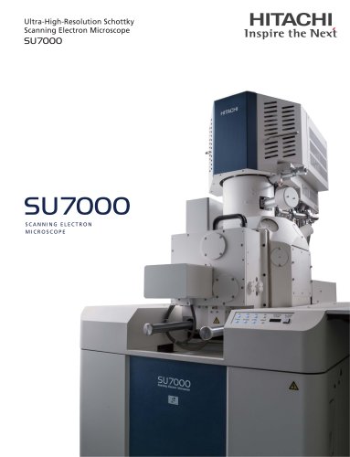
SU7000 SCANNING ELECTRON MICROSCOPE
1 /8Pages
Catalog excerpts
Ultra-High-Resolution Schottky Scanning Electron Microscope
Open the catalog to page 1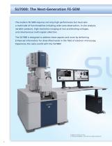
The modern FE-SEM requires not only high performance but must also a multitude of functionalities including wide-area observation, in-situ analysis, variable pressure, high-resolution imaging at low accelerating voltages, and simultaneous multi-signal collection. The SU7000 is designed to address these aspects and more by delivering Enhanced information for diversified needs in the field of electron microscopy. Experience the nano-world with the SU7000!
Open the catalog to page 2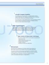
The SU7000 excels in fast acquisition of multiple signals to address expansive SEM needs, from imaging a wide field of view to visualizing sub-nanometer structures and everything in between. The incorporation of newly designed electron optics and detection systems allows for efficient simultaneous acquisition of multiple secondary electron and back-scattered electron signals. The number of the detectors mounted on the SEM is ever increasing, along with the need to display all collected information effectively. The SU7000 is capable of processing, displaying, and saving up to 6 signals simultaneously...
Open the catalog to page 3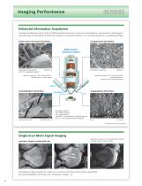
SID'7000 Enhanced information Acquisition The advanced detection system of the SU7000 streamlines acquisition of structural, topographical, compositional, crystallographic, and other types of information by minimizing changes to microscope conditions, such as working distance or accelerating voltage. SEM Column/ Detection System specimen courtesy of: Dr. Yoshiyuki Kubota Associate professor, Natural Science Research Institute Single-Scan Multi-Signal Imaging specimen: Organic-coated gold rods Simultaneous image acquisition for surface micro-structural information (UD), surface coating (MD), and...
Open the catalog to page 4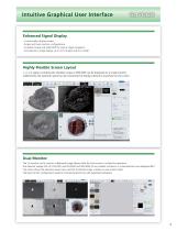
Intuitive Graphical User InterfaceEnhanced Signal Display •Customizable display modes. •Single and Dual-monitor configurations. •Chamber Scope and SEM MAP for optical stage navigation. •Simultaneous image display up to 4-ch (single) and 6-ch (dual). 1,2, or 4 signals including the chamber scope or SEM MAP can be displayed on a single monitor. Additionally, the operation panel can be customized to display submenus anywhere on the screen. Dual Monitor The 1st monitor can be used as a dedicated image display while the 2nd monitor is utilized for operation. Five detector images (UD, LD, UVD, MD,...
Open the catalog to page 5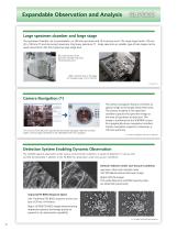
Expandable Observation and AnalysisLarge specimen chamber and large stage The specimen chamber can accommodate a cp 200 mm specimen and 18 accessory ports. The large stage travels 135 mm (X) x 100 mm (Y) and can accept maximum 2 kg heavy specimen.(*) Large specimen or variable type of sub-stages can be easily mounted on the front-opening large stage door. The camera navigation feature correlates an optical image to the target observation area. The camera installed in the specimen chamber captures the specimen image at the time of specimen introduction. The image is transferred to the SEM MAP...
Open the catalog to page 6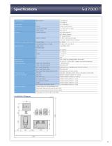
Specifications Image Resolution
Open the catalog to page 7
Notice: For correct operation, follow the instruction manual when using the instrument. Specifications in this catalog are subject to change with or without notice, as Hitachi High-Tech Corporation continues to develop the latest technologies and products for our customers. Copyright (C) Hitachi High-Tech Corporation 2022 All rights reserved. ^Hitachi High-Tech CorporationTokyo, Japanwww.hitachi-hightech.com/global/science/ Paper from “ responsible sources
Open the catalog to page 8All Hitachi High-Tech Europe GmbH catalogs and technical brochures
 HT7800 Series | TEM
2 Pages
 TM4000 Series
2 Pages
 VP (FE) SEM Family
4 Pages
Archived catalogs

- Automation software solution
- Hitachi laboratory microscope
- Inspection microscope
- Desktop microscope
- Digital camera microscope
- Measuring microscope
- Hitachi analysis microscope
- Industrial microscope
- Hitachi automated microscope
- Hitachi high-resolution microscope
- Bright field microscope
- Compact microscope
- Materials research microscope
- Metallurgical microscope
- Quality control microscope
- Research microscope
- Dark field microscope
- Ergonomic microscope
- Sample preparation system