- Products
- Catalogs
- News & Trends
- Exhibitions
Ultrasonic Nanoparticle Preparation for Pharmaceuticals
1 /7Pages

Ultrasonic Nanoparticle Preparation for Pharmaceuticals
1 /7Pages
Catalog excerpts

Ultrasonic Treatment of Nanoparticles for Pharmaceuticals Ultrasound is an innovative technology that is used successfully for sonochemical synthesis, deagglomeration, dispersion, emulsifying, functionalization and activation of particles. Particularly in nanotechnology, the ultrasonication is an essential technique for the synthesis and processing purposes of nano-size materials. Since nanotechnology has gained this outstanding scientific interest, nano-sized particles are utilized in extraordinarily many scientific and industrial fields. Also the pharma branch has discovered the high potential of this flexible and variable material. Consequently, nanoparticles are involved into various functional applications in the pharmaceutical industry, these include: • drug delivery (carrier) • diagnostic products • product packaging • biomarker discovery As nanomaterials are defined as particles with a dimension less than 100nm this means that the production and processing of these substances require higher efforts. Especially, drug delivery via nanoparticles is already a proven method for delivering active agents which have been administered before oral or by injection. (Bawa 2008) Nanoformulated drugs can be dosed and delivered much more efficient as new techniques open completely novel ways of medical treatments. This high-potential technology helps delivering drugs, heat, or other active substances to specific cells, i.e. diseased cells. By this direct drug delivery, healthy cells are untroubled by drug effects. One field, in that nanoformulated drugs already show their promising results is the cancer therapy. In the cancer therapy it is the big advantage of nano-sized substances that high doses of drug molecules can be delivered directly to the tumor cells for maximum effects while minimizing side effects to other organs. (Liu et al.2008) This advantage results in the nano-size by that the particles are able to pass cell walls and membranes and release the drug’s active agents directly at the targeted cells. To form and to process nanoparticles, agglomerates have to be broken and bonding forces have to been overcome. Ultrasonic cavitation is a well-known technology to deagglomerate and disperse nanomaterials. The diversity of nanomaterials and forms opens manifold changes for pharmaceutical research. Carbon Nanotubes (CNTs) have a large inner volume which allows more drug molecules to be encapsulated, and they have distinct inner and outer surfaces for functionalization. (Hilder et al. 2008) By that, CNTs are able to carry various molecules such as active agents, DNA, proteins, peptides, targeting ligands etc. into cells. CNTs have been recognized as the quintessential nanomaterials and have acquired the status of one of the most active fields of nanoscience and nanotechnology. The MWCNT is composed of 2–30 concentric graphitic layers, the diameters of which range from 10 to 50 nm and length more than 10 μm. On the other hand, SWCNT is much thinner, with diameter ranging from 1.0 to 1.4 nm. (Srinivasan 2008) Nanoparticles as well as nanotubes can enter cells and can be taken up by them completely. In particular funtionalized Carbon Nanotubes (f-CNTs) are known to enhance solubility and allow an efficient tumor targeting. By that, f-
Open the catalog to page 1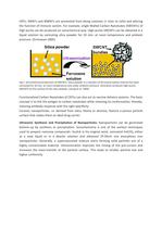
CNTs, SWNTs and MWNTs are prevented from being cytotoxic (= toxic to cells) and altering the function of immune system. For example, single-Walled Carbon Nanotubes (SWCNTs) of high purity can be produced on sonochemical way: High-purity SWCNTs can be obtained in a liquid solution by sonicating silica powder for 20 min. at room temperature and ambient pressure. (Srinivasan 2005) Fig.1: Sonochemical production of SWCNTs. Silica powder in a solution of ferrocene-xylene mixture has been sonicated for 20 min. at room temperature and under ambient pressure. Sonication produces high-purity SWCNTS on...
Open the catalog to page 2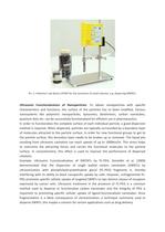
Pic. 1: Hielscher’s lab device UP50H for the sonication of small volumes, e.g. dispersing MWNTs. Ultrasonic Functionalization of Nanoparticles: To obtain nanoparticles with specific characteristics and functions, the surface of the particles has to been modified. Various nanosystems like polymeric nanoparticles, liposomes, dendrimers, carbon nanotubes, quantum dots etc. can be successfully functionalized for efficient use in pharmaceutics. In order to functionalize the complete surface of each individual particle, a good dispersion method is required. When dispersed, particles are typically surrounded...
Open the catalog to page 3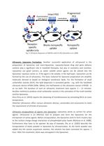
Fig. 2: Ultrasonic dispersion of SWCNTs with PL-PEG (Zeineldin et al. 2009) Ultrasonic Liposome Formation: Another successful application of ultrasound is the preparation of liposomes and nano-liposomes. Liposome-based drug and gene delivery systems play a significant role in manifold therapies, but also in cosmetics and nutrition. Liposomes are good carriers, as water soluble active agents can be placed into the liposomes’ aqueous center or, if the agent is fat soluble, in the lipid layer. Liposomes can be formed by the use of ultrasonics. The basic material for liposome preperation are amphilic...
Open the catalog to page 4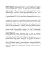
Liposomal Emulsions: To enhance the nurturing effect of moisturizing or anti-aging cremes, lotions, gels and other cosmeceutical formulations, emulsifier are added to the liposomal dispersions to stabilize higher amounts of lipids. But investigations had shown that the capability of liposomes is generally limited. With the addition of emulsifiers, this effect will appear earlier and the additional emulsifiers cause a weakening on the barrier affinity of phosphatidylcholine. Nanoparticles - composed of phosphatidylcholine and lipids – are the answer to this problem. These nanoparticles are formed...
Open the catalog to page 5
□ liposomes at 1 day afte r pre paration ■ liposomes a: 7 days alrer preparatio rT □ liposomes at 1 month after p reparation □ liposomes a: 2 m onths after preparadon ■ liposomes at 9 months after preparation □ liposomes a:' 2 months after prepa radon □ I rxs ;mes at ■ cay a'te" p 'enaraton ■ I pes ;mes si 7 days alter :repa a:t;r o liposomes at' month aner preparation □ liposomes at 2 months a'ter prepar-aopi ■ liposomes at & months a'ter preparaUsn □ liposomes at 12mon"Jis sper prepara-Jon Fig.l. Size distribution of MLV dispersions for 1 year. Fig.2. Size distribution of SUV dispersions for...
Open the catalog to page 6All Hielscher catalogs and technical brochures
 Ultrasonic Process Lab
1 Page
 Hielscher Ultrasonic Sieves
2 Pages
 Ultrasonic Wire Cleaning
4 Pages
 Ultrasonic Applications
1 Page


- Liebherr laboratory stirrer
- Homogenizer
- Liebherr analog laboratory stirrer
- Liebherr beaker laboratory stirrer
- Liebherr mechanical laboratory stirrer
- Liebherr compact laboratory stirrer
- Batch homogenizer
- Liebherr overhead laboratory stirrer
- Sieve shaker
- Liquid homogenizer
- Liebherr vertical laboratory stirrer
- Liebherr disperser
- Dry sieving sieve shaker
- Vertical homogenizer
- Laboratory homogenizer
- Wet sieving sieve shaker
- Liebherr laboratory disperser
- Automatic homogenizer
- Ultrasonic homogenizer
- Laboratory sieve shaker