
- Catalogs
- Evident - Olympus Scientific Solutions
- GX53 Brochure


- Company
- Products
- Catalogs
- News & Trends
- Exhibitions
GX53 Brochure

GX53 Brochure
The GX53 inverted metallurgical microscope is designed for industries such as steel, automotive, and electronics. It allows inspection of polished metals and cross-section samples without leveling, accommodating thick, large, or heavy samples. The microscope provides high-quality images and, when paired with PRECiV™ software, enhances the inspection process from observation to reporting.
- Quick Analysis: Enables rapid observation, measurement, and analysis of metallurgical structures.
- Advanced Imaging: MIX technology combines darkfield with other methods to enhance sample surface visibility.
- HDR and EFI: High dynamic range (HDR) and extended focus imaging (EFI) improve image clarity and focus.
- Particle and Grain Analysis: PRECiV software supports particle analysis and grain sizing, adhering to various industrial standards.
- MIX Technology: Combines brightfield and darkfield for unique observation images, useful for visualizing surface textures.
- HDR: Adjusts brightness differences to reduce glare and enhance contrast.
- EFI: Stacks images to create all-in-focus images, even for uneven surfaces.
The GX53 and PRECiV software support metallurgical analysis methods compliant with standards like ISO, ASTM, DIN, and JIS. The software provides step-by-step guidance for efficient analyses.
The ergonomic design of the GX53 ensures user comfort, with features like a tilting observation tube and convenient hand switch. The microscope supports multiple observation methods and allows easy switching between them.
PRECiV software simplifies report creation with predefined templates and coded hardware settings for consistent inspections. Auto calibration and shading correction enhance measurement reliability and image quality.
The GX53 features high-intensity white LED illumination for consistent color temperature and reliable image quality. The MXPLFLN objectives offer high numerical aperture and long working distance, improving inspection speed and accuracy.
The GX53 is customizable with various optical components and PRECiV image analysis packages to meet specific inspection needs.
The GX53 microscope utilizes the UIS2 optical system, which is infinity-corrected. It features manual brightfield/darkfield selection by mirror unit and a manual field stop/aperture stop switch with centering. Light source options include a white LED with a Light Intensity Manager, a 12V 100W halogen lamp, a 100W mercury lamp, and a light guide source. Supported observation modes are brightfield, darkfield, differential interface contrast (DIC), simple polarizing, and MIX observation with 4 directional darkfield.
Various scale glasses are available for different magnifications: 10× (100 μm), 20× (50 μm), 50× (10 μm), and 100× (10 μm). Additionally, a grain size scale is applicable to standards such as JIS G 0551, ISO 643, and ASTM E112.
The GX53 microscope includes a built-in power supply for reflected light and a camera adaptor port for displaying live and captured images. Users can choose from various light sources and power supplies, including standard LED, high-intensity, and halogen configurations.
Adaptors are available for adding cameras, with options based on field of view and magnification. The actual observation range can be calculated using a specific formula.
Various tubes and eyepieces are available for imaging through eyepieces or cameras, with options for ergonomic comfort and imaging type.
Nosepieces are used to attach objectives and sliders, with options for brightfield, darkfield, and MIX observation. Sliders provide topographic information and illumination flexibility.
Control boxes connect the microscope’s hardware with a PC, and hand switches are available for hardware display and control.
Stages and stage plates are available for sample placement, with options based on sample shape and size.
Optical filters convert sample exposure light to various types of illumination, with options for different observation requirements.
Objectives magnify the sample, with options based on working distance, resolving power, and observation method. Various types are available, including brightfield, darkfield, and polarizing objectives.
The GX53 system diagram outlines the configuration of camera adaptors, tubes, eyepieces, frames, illuminations, nosepieces, and other components, providing a comprehensive overview of the system setup.
The document outlines specifications for a digital microscope system, including a 5 V DC, 2.5 A power input with an AC adaptor compatible with 100–240 V, 0.4 A, 50/60 Hz. It features transmitted light illumination requiring an optional power supply, and a continuously-variable light intensity dial by voltage. The external interface includes connectors for a coded nosepiece, MIX Slider, handsets, RS-232C, and USB 2.0.
The focus system uses a rack and pinion with a roller guide, offering manual coarse and fine coaxial handling. The focus stroke is 9 mm, with fine handle stroke per rotation at 100 μm and coarse handle stroke at 7 mm. It includes a torque adjustment ring and an upper limit stopper for coarse focusing.
The microscope supports widefield inverted tubes and a nosepiece with 4 to 7 holes for brightfield and 5 to 6 holes for brightfield/darkfield, with manual or coded types and optional centering.
The stage options include a right handle stage for GX with X/Y stroke of 50 × 50 mm and a max load of 5 kg, and flexible right and left short handle stages with a max load of 1 kg.
The microscope is designed for indoor use with an ambient temperature range of 5 to 40 °C and a maximum relative humidity of 80% up to 31 °C. It adheres to pollution degree 2 and installation category II standards.
The document mentions the DSX1000 Digital Microscope and LEXT OLS5100 Laser Scanning Microscope, highlighting their precision and efficiency in measuring shape and surface roughness at the submicron level.
Evident Corporation is ISO14001 and ISO9001 certified. The product is designed for industrial environments and may affect other equipment if used in residential settings. Periodic inspections of illumination devices are recommended.
For more details, the document directs readers to visit the Olympus-IMS website.
Catalog excerpts

GX53 Inverted Metallurgical Microscope Advanced Microscopy Solutions for Metallurgy Inspection
Open the catalog to page 1
Quick Analysis for Large or Thick Samples The GX53 inverted microscope is used for a wide range of applications often seen in the steel, automotive, electronics, and other manufacturing industries. The microscope enables users to inspect polished metals and cross-section samples simply by placing them upside down on the stage. The sample does not need to be leveled and can be thick, large, or heavy. The GX53 delivers crisp images that can be difficult to capture using conventional microscopy observation methods. When combined with PRECiV ™ image analysis software, the microscope streamlines the...
Open the catalog to page 2
Streamline Your Inspection Process Fast Inspections, Advanced Functionality Quickly observe, measure, and analyze metallurgical structures. User Friendly Even novice operators can comfortably make observations, analyze results, and create reports. Advanced Imaging Technology Our proven optics and imaging technology deliver clear images and reliable results. Modular Choose the components you need for your application.
Open the catalog to page 3
Fast Inspections, Advanced Functionality Advanced Analysis Tools The GX53 microscope’s various observation capabilities provide clear, sharp images, so users can reliably detect defects in their samples. PRECiV™ image analysis software's illumination techniques and image acquisition options give users more choices for evaluating their samples and documenting their findings. The Invisible Becomes Visible: MIX Technology MIX technology produces unique observation images by combining darkfield with another observation method, such as brightfield or polarization. MIX observation enables users to...
Open the catalog to page 4
Easily Create Panoramic Images: Instant MIA Create All-in-Focus Images: EFI With multiple image alignment (MIA), users can stitch images together quickly and simply by moving the XY knobs on the manual stage—a motorized stage is optional. PRECiV ™ software uses pattern recognition to generate a panoramic image, which is suitable for inspections of carburizing and metal-flow conditions. PRECiV software’s extended focus imaging (EFI) function captures images of samples whose height extends beyond the depth of focus. EFI stacks these images together to create a single all-in-focus image of the sample....
Open the catalog to page 5
PRECiV™ Software—Optimized for Materials Science Material inspection, measurement, and analysis are required to comply with industrial standards as well as internal operating procedures. Together, the GX53 microscope and PRECiV software support metallurgical analysis methods that comply with different industrial standards. With step-by-step operator guidance, users can analyze their samples quickly and easily. Particle Analysis—Count and Measure Solution Detecting objects and measuring size distribution are among the most important applications in digital imaging. PRECiV software’s Count and...
Open the catalog to page 6
Evaluating Graphite Nodularity The software can be used to evaluate the graphite nodularity and content in cast iron samples (nodular and vermicular). The form, distribution, and size of graphite nodes can be classified. Supported standards: ISO, NF, ASTM, KS, JIS, GB/T Ductile cast iron showing nodular graphite Rating Nonmetallic Inclusion Content in High-Purity Steel Classify nonmetallic inclusions using an captured image of the worst field or inclusion that you have manually located on the sample. Supported standards: ISO, EN, ASTM, DIN, JIS, GB/T, UNI Steel with nonmetallic inclusions Compare...
Open the catalog to page 7
User Friendly A Design That Emphasizes User Comfort The microscope’s ergonomic design helps users stay comfortable while they work, contributing to a more efficient inspection. When used with PRECiV™ software, operators can easily acquire images of diverse samples, conduct a variety of analyses, and generate professional reports. Maintain a comfortable posture The tilting observation tube’s extensive range and adjustable eyepoint enable operators to sit or stand at the microscope in a comfortable posture. Observe large, heavy samples Samples up to 5 kg (11 lb) can be inspected simply by placing...
Open the catalog to page 8
Easily Restore Microscope Settings: Coded Hardware Coded functions integrate the microscope’s hardware settings with PRECiV™ image analysis software. The observation method, illumination intensity, and magnification can be recorded by the software and stored with the associated images. Since the settings can easily be reproduced, different operators can conduct the same quality inspections with limited training. Retrieve the device settings with PRECiV software Operator B Restore Device Status menu All operators can use the same settings. Different operators use different settings. User Guidance...
Open the catalog to page 9
Proven Optics and Digital Imaging Technology Deliver Quality Data Our history of developing high-quality optics and advanced imaging capabilities has led to quality microscopes that offer exceptional measurement accuracy. Reliable Optical Performance: Wavefront Aberration Control The optical performance of objective lenses directly impacts the quality of the observation images and analysis results. Our UIS2 high-magnification objectives are designed to minimize wavefront aberrations, delivering reliable optical performance. Consistent Color Temperature: High-Intensity White LED Illumination The...
Open the catalog to page 10
Applications Reflected light microscopy spans a range of applications and industries. Below are just a few examples of what can be achieved using different observation methods. Polished sample of AlSi Spheroidal graphite cast iron Brightfield is a common observation method to observe reflected light from a sample by illuminating it straight on. Darkfield is used to observe scattered or diffracted light from a sample, so imperfections clearly stand out. Inspectors can identify even minute scratches or flaws. Differential interference contrast (DIC) is an observation technique where the height...
Open the catalog to page 11All Evident - Olympus Scientific Solutions catalogs and technical brochures
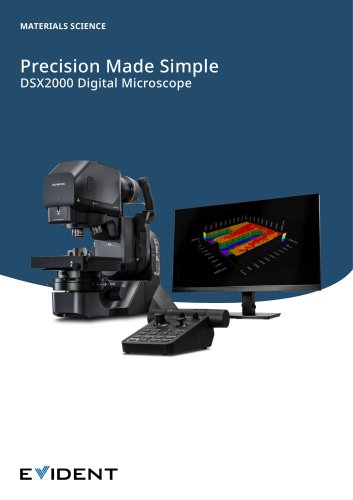 DSX2000 Digital Microscope
15 Pages
 MXPLFLN Series
2 Pages
 OLYMPUS Stream
16 Pages
 LC35
LC354 Pages
 DP74
DP748 Pages
 SZX7
SZX716 Pages
 SZ61 SZ51 Brochure
24 Pages
 SZX16 - SZX10
24 Pages
 MX63/MX63L
20 Pages
 BXC Series Brochure
4 Pages
 BX53M - BXFM Brochure
28 Pages
 CIX100
16 Pages
 STM7 Series
24 Pages
 LEXT OLS5000
42 Pages
 Camera Overview
12 Pages
 DSX1000 Brochure
40 Pages
 PRECiV 1.2 Brochure
20 Pages
 SZX-AR1
12 Pages
- Digital imager
- Visible imager
- CMOS camera module
- Automation software solution
- Full-color camera system
- Control software
- USB camera module
- EVIDENT optical microscope
- Industrial software
- Measurement software
- Inspection imager
- HD camera module
- Laboratory microscope
- Quality software
- USB 3.0 camera module
- Inspection microscope
- Desktop microscope
- LED illumination microscope