 Website:
Bruker AXS
Group: Bruker Corporation
Catalog excerpts

Technology Guide D8 DISCOVER Diffraction Solutions Innovation with Integrity
Open the catalog to page 1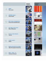
Dedicated hardware components XRD – True musts DAVINCI design – Fully loaded Don’t miss a single phase – Phase ID & Quantification Get right to the point – Micro Diffraction Focus on the relevant – Texture Under control – Stress Master the flood of samples – High-Throughput Screening Micro – Nano – Å – Structure Analysis
Open the catalog to page 2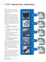
XRD - discover the g-information XRD – X-ray diffraction by means of a 2-dimensional detector – is the method of choice for examining any type of sample in a nondestructive way. An XRD pattern covers a large solid angle containing several complete or large Single Crystal portions of Debye rings that describe the diffracted intensity distribution in both 2θ and γ directions. An XRD pattern therefore contains abundant information about the properties of a crystalline material. Extracting this information is straightforward using our comprehensive XRD algorithms. Here’s Micro Sample how it...
Open the catalog to page 3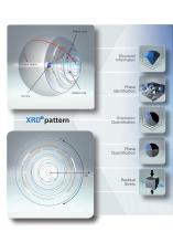
Debye cone Structural Information Incident beam Debye ring Phase Identification Orientation Quantification Phase Quantification Residual Stress
Open the catalog to page 4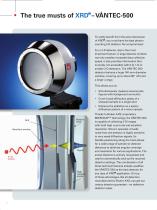
To really benefit from the extra dimension of XRD, you must have the best photon counting 2-D detector. No compromises! For a 2-D detector, size is the most important feature. A large detector window not only enables increased data collection speed, it also provides information that is simply not accessible with 0-D, 1-D or smaller 2-D detectors. The VÅNTEC-500 detector features a huge 140-mm-diameter window, covering up to about 80° (2θ) and a large γ-range. This allows you to: imultaneously measure several pole S figures with background correction over broad diffraction peaks of a C...
Open the catalog to page 6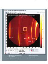
XRD pattern of a textured Cu thin film with VÅNTEC-500 in 1:1 scale Size of standard solid-state 2-D detector uge detection area covering more H than 15,000 mm² ariable detector positioning with V real-time distance recognition Maintenance-free design uaranteed to have no dead G or defective areas
Open the catalog to page 7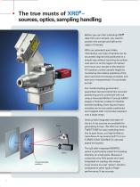
The true musts of XRD – sources, optics, sampling handling Before you can start collecting XRD data from your sample, you need to position the sample and define the region of interest. With our patented Laser-Video microscope, any type of sample can be accurately aligned and positioned in a simple way without touching its surface. Just zoom in on the region of interest and move your sample to the desired XY-position; control sample height by monitoring the relative positions of the laser spot and microscope crosshair and start your measurement. It is as simple as that. Our market-leading...
Open the catalog to page 8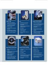
■ Fast and easy switching between line and spot focus ■ Compatible with standard sealed tube dimensions ■ Automatic focus orientation disconnect cables and hoses High-Precision Goniometer ■ Vertical or horizontal goniometer, ■ Stepper motors with optical encoders ■ Fast and reliable positioning ■ Dovetail tracks for flexible ■ Microfocus X-ray source ■ Fully integrated into the ■ With integrated parallel-beam MONTEL mirror Flexible Sample Handling ■ XYZ stages for large and heavy ■ Sample spinners for capillaries and flat powder samples TURBO X-RAY SOURCE ■ Rotating anode generator focus...
Open the catalog to page 9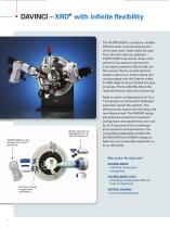
DAVINCI – XRD with infinite flexibility The D8 DISCOVER is a uniquely modular diffractometer, incorporating all parts of the beam path. Easily select the tube focus direction with our patented TWIST-TUBE X-ray source. Snap in the desired X-ray optics to optimize the X-ray beam properties. Mount add-ons like vacuum chucks, sample spinners, capillary spinners or dome heating and cooling stages onto the Eulerian cradle or UMC stage to accommodate any type of sample. Put the VÅNTEC-500 at the required distance, then start measuring. Widest selection of dedicated 0-D, 1-D and 2-D detectors...
Open the catalog to page 10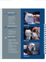
DAVINCI.MODE – real-time component recognition nstant component I registration with all specific properties ail-safe component F positioning True plug’n’play utomatic detector A distance recognition DAVINCI.SNAP-LOCK – changing components without tools Fast and easy lignment-free: optics A retain their alignment ariable positioning V on the track DIFFRAC.DAVINCI – the virtual goniometer eal-time component R recognition and status display ush-button switch between P high-resolution and high-flux beam path etection of missing, D misplaced or unsuitable components hoice between all C...
Open the catalog to page 11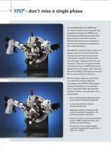
XRD – don’t miss a single phase The D8 DISCOVER with XRD each and every sample can be analyzed. The exceptional strength of XRD excels at analyzing challenging samples with larger grains or textured materials. Samples like these can be analyzed in real time at dimensions unmatched by other diffractometers. Preparation is quick and easy: place your sample, zoom in with the Laser-Video microscope and center your sample – nothing else required. Choose a start and end angle, measurement time, and resolution. Then go! The system starts collecting a series of XRD patterns and displays them in a...
Open the catalog to page 12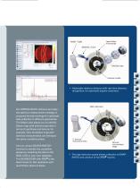
Laser-Video microscope TWIST - TUBE MONTEL mirror Eulerian cradle Adjustable detector distance with real-time distance recognition, for optimized angular resolution. Our DIFFRAC.SUITE software provides the optimum measurement strategy. It proposes the best settings for optimized data collection in different geometries. The Debye view allows you to identify Debye rings with similar properties in terms of spottiness and texture, for example. Only the Debye rings with identical characteristics can belong to the same crystalline phase. Use our unique SEARCH/MATCH function to identify the...
Open the catalog to page 13
XRD – get right to the point Patterned wafers, forensic samples, inclusions in geological materials… These very diverse samples all have one thing in common: the area of interest is very tiny. For these applications, our patented Laser-Video microscope and precise sample stages guarantee that you exactly measure the area of interest–regardless of sample size or shape. When an X-ray beam is collimated down to a micro-spot size, obviously only a few crystallites are hit by the incident beam and diffract the X-rays. This results in spotty diffraction patterns and in very weak diffraction...
Open the catalog to page 14All Bruker AXS catalogs and technical brochures
-
XRF SPECTRA ELEMENTS
4 Pages
-
D8 DISCOVER Plus
4 Pages
-
S2 LION - Spectrometry Solutions
12 Pages
-
S2 PUMA - Spectroscopy Solutions
20 Pages


