- Catalogs
- Bruker AXS
- Micro-CT for SEM - Nondestructive Measurement and Volume Visualization of Specimens`Internal Microstructure in SEM
- Company
- Products
- Catalogs
- News & Trends
- Exhibitions
Micro-CT for SEM - Nondestructive Measurement and Volume Visualization of Specimens`Internal Microstructure in SEM
1 /8Pages

Micro-CT for SEM - Nondestructive Measurement and Volume Visualization of Specimens`Internal Microstructure in SEM
1 /8Pages
Catalog excerpts

Micro-CT for SEM Nondestructive Measurement and Volume Visualization of Specimens’ Internal Microstructure in SEM Innovation with Integrity
Open the catalog to page 1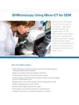
3D Microscopy Using Micro-CT for SEM Micro-CT for SEM adds true 3D microscopy to your SEM – regardless of manufacturer and model. Micro-CT for SEM extends the surface information gained with conventional SEM imaging by allowing an unique insight into the specimen’s internal microstructures – nondestructively and with ease of use. Obtain information on a specimen’s internal microstructure nondestructively and without any additional sample preparation Measure and visualize the internal morphology in 2D and 3D Generate realistic models for a virtual travel through a specimen Intuitive and easy-to-use...
Open the catalog to page 2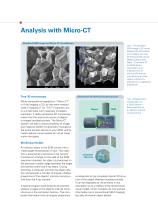
Analysis with Micro-CT Standard SEM image and Micro-CT visualization True 3D microscopy Micro computed tomography or “Micro-CT” is X-ray imaging in 3D, by the same method used in hospital CT (or “CAT”) scanners, but on a small scale with massively increased resolution. It really represents 3D microscopy, where the fine internal structure of objects is imaged nondestructively. The Micro-CT system will add a unique possibility to image and measure 2D/3D morphometry throughout the entire sample volume to your SEM, and to create realistic visual models for virtual travel within the object. Left –...
Open the catalog to page 3
Microscanner and X-ray camera The Micro-CT system for SEM contains several modules to add a 3D imaging modality to any SEM without compromising standard imaging modes. The Micro-CT system does not require any changes to the standard SEM construction. A microscanner contains the target to produce X-rays, an object rotation stage, and a motorized linear stage to vary the distance between the X-ray emission point and the object for adjusting the magnification of the X-ray images. The microscanner can be installed inside the specimen chamber in place of standard object holders. A camera assembly...
Open the catalog to page 4
A Wide Field of Application Composite materials Glass fiber/epoxy composite material containing 10–12 micron fibers in an epoxy matrix. From left to right: 1) SEM image in SE mode; 2) X-ray image through this sample, acquired using the Micro-CT for SEM; 3) a virtual section, obtained nondestructively by Micro-CT for SEM; 4) a 3D model of the internal microstructure built from all reconstructed cross sections with a virtual cut on an inclined plane; all X-ray images have a pixel size of 705 nm Heat shielding ceramic Top left – SEM image in SE mode Top right – X-ray shadow image Bottom left – a...
Open the catalog to page 5
Filter inspection Used filter from a vacuum cleaner filled with dust particles. From left to right: 1) SEM image of the filter surface in SE mode. The other three images show the results of Micro-CT reconstruction where the filter material is shown in silver, and dense particles in red; 2) front view; 3) back view; 4) side view with semitransparent filter material; notice that most particles are absorbed within the front surface of the filter (figures 2 and 4) and cannot pass through the filter material Wood and plants Top – SEM image of wood sample in SE mode Right – orthogonal virtual slices...
Open the catalog to page 6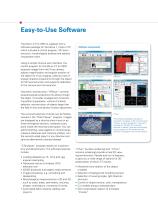
Easy-to-Use Software The Micro-CT for SEM is supplied with a software package for Windows 7, Vista or XP, which includes a control program, 3D reconstruction, morphological analysis and realistic visualization tools. Using a simple intuitive user interface, the control program for the Micro-CT for SEM acquires images from the X-ray camera, adjusts magnification and angular position of the object for X-ray imaging, collects a set of angular shadow projections through the object for 3D reconstruction and supports calibration of the camera and microscanner. Volumetric reconstruction “NRecon” converts...
Open the catalog to page 7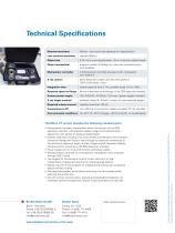
Technical Specifications Nominal resolution 400 nm – 8 µm pixel size (depends on magnification) Low-contrast resolution Object size 0.18 – 4 mm scanning diameter, 10 mm maximum object length Object manipulator precision rotation 0.45 deg min. step size; motorized zoom with feedback Manipulator controller microprocessor controller powered (with manipulator) from USB 2.0 X-ray camera direct detection cooled CCD, 512 x 512 pixels or 1024 x 1024 pixels, 16-bit Integration time typical exposure time: 2 – 4 s, possible range: 0.5 to >100 s Required space on flange 65 mm in diameter on any flange of...
Open the catalog to page 8All Bruker AXS catalogs and technical brochures
 XRF SPECTRA ELEMENTS
4 Pages
 D8 DISCOVER Plus
4 Pages
 S2 LION - Spectrometry Solutions
12 Pages
 S2 PUMA - Spectroscopy Solutions
20 Pages


- Analysis software solution
- Microscope
- Bruker spectrometer
- 3D software solution
- Visualization software solution
- Laboratory microscope
- Bruker laboratory spectrometer
- 2D software
- Inspection machine
- Desktop microscope
- Compact spectrometer
- Process spectrometer
- Benchtop spectrometer
- High-resolution microscope
- Analysis spectrometer
- Optical spectrometer
- Compact microscope
- Measurement spectrometer
- Portable spectrometer
- High-resolution spectrometer