BC43
1 /13Pages

BC43
1 /13Pages
Catalog excerpts

BC43 The Ultimate Benchtop Confocal Microscope Key Features Benchtop multimodal imaging system Instant confocal: Blur-free imaging Widefield imaging Differential phase contrast & brightfield Borealis uniform illumination GPU-powered deconvolution Cell biology Developmental biology Neuroscience Cancer biology Tissue imaging Organoids & large organisms
Open the catalog to page 1
Introduction | Applications | Software | Features | Specifications | Ordering Andor Benchtop Confocal 2D and 3D imaging as easy as ABC Advanced imaging technology Sharp 2D & 3D images instantly. Enhanced visualisation software Intuitive and powerful. Achieve outstanding results quickly with minimal training. Easy to use Ergonomic joystick and 2x objective allow quick sample overview. Benchtop design Light tight lid and inbuilt anti-vibration, so no need for a darkroom or optical table. Optimal performance Multidimensional experiments possible. Patented Focus Seek & Lock ensures accuracy in acquisition....
Open the catalog to page 2
Introduction | Applications | Software | Features | Specifications | Ordering Andor Benchtop Confocal Total Imaging Flexibility Confocal Imaging Confocal technology provides high-contrast, blur-free images. It boosts image quality of thin samples, such as monolayer cultures, and is especially suited for thick samples like small model organisms, 3D cultures and cleared tissues. Widefield & Deconvolution BC43 captures images at least 10x faster than point scanning confocals, boosting productivity, yet maintaining full resolution. Image deeper with higher quality than solutions that rely on computational...
Open the catalog to page 3
Introduction | Applications | Software | Features | Specifications | Ordering “I found BC43 super easy to setup Application Focus for all my experiments and super fast to acquire and deliver highquality data. I love its flexibility.” Developmental Biology Marco Campinho, BC43 cuts through the challenges easily, spanning development from the first rounds of cell division to the fully developed organism. Use BC43 to image at depth, in gentle live imaging experiments of cells and tissues. Effortlessly acquire multiple Z stacks, multiple tiles in combination with time-lapse imaging. Group Leader...
Open the catalog to page 4
Introduction | Applications | Software | Features | Specifications | Ordering Application Focus Cell Biology Working closely with leading cell biologists we have carefully developed BC43 to meet the needs of a broad range of experiments. Reveal the detail inside cells from nm to mm within tissues and whole model organisms with BC43. Use BC43 in confocal mode to see detail hidden in the sample background or image in widefield to increase sensitivity and speed. Image fast dynamic events, such as microtubule dynamics, or study longer processes like cell cycle over 24 hours with no photobleaching...
Open the catalog to page 5
Introduction | Applications | Software | Features | Specifications | Ordering Application Focus Tissue Imaging Large area imaging needs to provide both cellular resolution and the full organ context. The advanced high-speed technology in BC43 means you no longer need to compromise. Large area tissue confocal imaging is now possible. Ten times faster than regular confocals. No sacrifices in resolution, or field of view. BC43 delivers results fast, shortening the time to publication. Discover more in intact tissues, use cleared samples and BC43 in confocal mode to image even thicker samples. BC43...
Open the catalog to page 6
Introduction | Applications | Software | Features | Specifications | Ordering Application Focus Neuroscience BC43 is the perfect workhorse for neuroscience. Imaging experiments commonly require high magnification, for resolution, imaging of large areas to fully understand the architecture and connectivity of this complex tissue. The incredible confocal capture rate of BC43 dramatically reduces imaging time delivering results faster. BC43 is a push-button confocal suitable for the broadest range of cancer experimental models. Capturing stunning images of: • Subcellular events (e.g. cytoskeletal...
Open the catalog to page 7
Introduction | Applications | Software | Features | Specifications | Ordering Integrated Software Solutions Small in size, Big in performance BC43 is an ideal instrument for a core facility, easy to operate, with multiple microscopy techniques. It provides great images fast, whatever the sample. Free up your more complex imaging systems for users doing highly specialised experiments. BC43 has an integrated, easy-to-use, and accessible software interface that delivers high-end imaging. Users benefit from easy protocol set up for multidimensional experiments, such as oneclick multi–position-montage...
Open the catalog to page 8
Introduction | Applications | Software | Features | Specifications | Ordering Simple Workflows Fast to learn & time saving Here we show two possible workflows. All options can be performed in combination. Select the area of sample to be imaged. Select required objective. Set centre of Z scan. Press Acquire. Snap or overview the sample and move to the desired objective. Select the positions to be imaged. Press Acquire.
Open the catalog to page 9
Introduction | Applications | Software | Features | Specifications | Ordering Key Features of BC43 Hardware Feature Software Feature High-speed confocal imaging 3 3D optical sectioning with high background rejection. Eliminates blur. 3 Allows deep and large tissue imaging at speed for higher productivity. 3 Image fast dynamic events in thicker samples. 3 Fast to learn and easy-to-use multidimensional acquisition software. 3 Integrated confocal, widefield and brightfield imaging options. 3 Post-acquisition processing with stitching and deconvolution. Widefield imaging 3 Image thin specimens/structures...
Open the catalog to page 10
Introduction | Applications | Software | Features | Specifications | Ordering Mechanical Drawings Microscope Unit BC43 Imaging Modes Imaging Methods ClearView™ GPU High-speed confocal Widefield epifluorescence Transmitted light - brightfield and Differential Phase Contrast 2 Units: Millimeters [Inches] Single colour, multicolour, z-stacking (volume), time-lapse, multi-position, multi-well, montage and 2/3D stitching. A Clears image of non-specific sample background signal and improves resolution beyond A the normal optical limits. 6.5 μm pixel; 2048x2000 pixels (4.1 MP) Up to 82% 18.4 mm (diagonal)...
Open the catalog to page 11All Andor Technology catalogs and technical brochures
 Marana sCMOS
9 Pages
 MicroPoint 4
9 Pages
 ZL41 Cell sCMOS
7 Pages
 Sona sCMOS
9 Pages
 Optistat
11 Pages
 Multi-Wavelength Imaging
11 Pages
 andor-kymera-193-specifications
15 Pages
 andor-dragonfly-specifications
14 Pages
 Solis-Brochure
6 Pages
 Spectroscopy-Solutions-Brochure
24 Pages
 iKon-M/L SO Series
10 Pages
 iStar CCD and sCMOS
8 Pages
 Neo 5.5 sCMOS
6 Pages
 Mechelle 5000
5 Pages
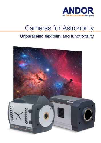
 Cameras for Astronomy
8 Pages
 Microspectroscopy Gatefold
8 Pages
 Spectroscopy Brochure
25 Pages
 iXon EMCCD
13 Pages
 Zyla for Physical Sciences sCMOS
12 Pages
 iVac OEM 2
2 Pages
 iKon-M OEM 2-page PV
2 Pages
 iKon-M X-Ray 2
2 Pages
 Newton EMCCD
16 Pages
 sCMOS
sCMOS16 Pages
 Darkcurrent
3 Pages
 iXon Back-illuminated
6 Pages
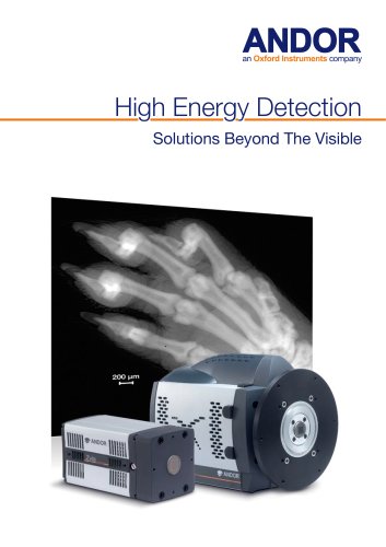 High Energy Detection_2014
28 Pages
 PRODUCT PORTFOLIO 2013
51 Pages
 Active Illumination Solutions
25 Pages
 Neo and Zyla sCMOS
27 Pages
 iXon
iXon33 Pages


Archived catalogs
 Intensified Camera Series
9 Pages
 Clara Interline CCD Series
2 Pages
 Andor Revolution XD brochure
23 Pages
 Revolution DSD
11 Pages
 Clara Flyer
2 Pages
 Revolution
23 Pages
 Low Light Imaging
8 Pages
 iQ Software
12 Pages
 Luca vs Interline CCD
6 Pages
 ApogeeAspen CG9000
5 Pages
 Apogee Aspen CG6
5 Pages
 Apogee Aspen CG47
5 Pages
 Apogee Aspen CG230
5 Pages
 Apogee Alta F9000
5 Pages
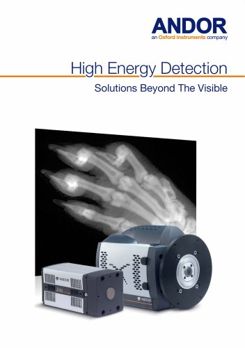 High Energy Detection_2019
8 Pages
 Astronomy Brochure_2014
19 Pages
 Astronomy Brochure
19 Pages
 iKon-M USB X-Ray Brochure
2 Pages


- Bourn And Koch digital camera
- Bourn And Koch visible camera
- Bourn And Koch management software
- Bourn And Koch automation software
- Bourn And Koch CMOS camera
- Bourn And Koch industrial camera
- Bourn And Koch analysis software
- Infrared imager
- Bourn And Koch process software
- Monitoring camera system
- Bourn And Koch full-color camera
- Bourn And Koch Windows software
- Bourn And Koch real-time software
- Bourn And Koch cloud software
- Bourn And Koch control software
- Bourn And Koch spectrometer
- Bourn And Koch USB camera
- Bourn And Koch detection camera
- Bourn And Koch image processing camera