- Catalogs
- Andor Technology
- Active Illumination Solutions
Active Illumination Solutions
1 /25Pages

Active Illumination Solutions
1 /25Pages
Catalog excerpts

Active Illumination Solutions Observe, Perturb, Measure
Open the catalog to page 1
Since the acquisition of Photonic Instruments in 2010 Andor has built on an existing FRAPPA offering to provide a comprehensive Active Illumination portfolio. Photonic Instruments was founded in 1996 and grew to be a market leader in Active Illumination and laser ablation systems for confocal and widefield microscopy. Andor has continued to enhance product performance and features, as well as expanding the range of available products.
Open the catalog to page 2
WHAT IS ACTIVE ILLUMINATION? Active Illumination, or AI, describes a rapidly evolving range of optical techniques with an increasing impact on scientific enquiry and experimentation. AI has developed over the last two decades alongside the revolution of fluorescent proteins in biology (ref 7, 8), the instrumental and technological developments of confocal laser scanning microscopy (CLSM), solid state light sources (lasers and LEDs), fast galvo and optical MEMS technology, and of course the ubiquitous personal computer. Many of these techniques were first envisaged or implemented in CLSM, but...
Open the catalog to page 3
WHAT IS ACTIVE ILLUMINATION? Uncaging Of Caged Compounds Caged compounds are light-sensitive probes that functionally encapsulate biomolecules to render them inactive. Targeted illumination releases the biomolecule enabling localized perturbation of biological processes. Caged compounds are commonly released with UV light and when used with fluorescence microscopy provide a powerful tool for observation, perturbation and measurement. Photoactivation Photoactivatable fluorescent proteins can undergo dramatic changes in their properties in response to absorption of specific illumination. They can...
Open the catalog to page 4
MicroPoint Simultaneous and precise illumination and ablation MicroPoint provides a flexible and field-proven tool for photo-stimulation. Supplied with a patented compact, pulsed nitrogen pumped tunable dye laser it is capable of ablation, bleaching and uncaging over a wavelength range of 365 to 656 nm. Broad wavelength range and energy control allow MicroPoint to be optimized for a wide range of scenarios. More than 20 wavelengths can be utilized with available dye resonator cells, while appropriate dichroic filter sets enable simultaneous imaging and photo-stimulation of the specimen. MicroPoint...
Open the catalog to page 5
MICROPOINT Configuring MicroPoint to your exact needs Dye Cells & Laser Dye MicroPoint is a highly versatile illumination source, compatible with all leading microscopes and most legacy systems. There are a selection of control interfaces, filter and laser/ lamp attachment options. Control Interface Galvo Bluetooth UV - 365 nm Dye Cell (Supplied with) Multiline Dye Cell (Supplied with) High Power 435 nm Dye Cell (Supplied with) Control Interface Pulse Generator (optional) Leica Other Laser Pulsed Nitrogen Laser System Includes 2M delivery fibre, Microscope specific interlock and Microscope Epi...
Open the catalog to page 6
TYPICAL APPLICATIONS Due to the broad range of wavelengths MicroPoint can be tuned to, it is used for a broad variety of applications, popular with core facilities. The more frequent applications are ablation (365 nm) in fields such as developmental biology and DNA damage. Both of these applications take advantage of the diffraction limited spot to target single cells, or subcellular components. Diffraction Limited FRAP Yeast cells are labeled with Nuf2p: GFP to mark the spindle pole bodies and Ase1p: GFP in the mitotic spindle mid-zone. They are observed in DIC and fluorescence microscopy. Ablation...
Open the catalog to page 7
Share all laser lines for photo stimulation and imaging “Bypass” mode provides 1:1 relay imaging for “in-line” configuration FRAPPA is a galvo scanning instrument, named by conjoining acronyms for fluorescence recovery after photobleaching (FRAP) and photoactivation (PA). FRAPPA has a unique switching design that allows it to be configured in the imaging path. In bypass mode it acts as a relay optic, projecting an image to the detector; while in scanning mode it acts as a laser scanner, targeting user-defined regions of the specimen. This “in-line” configuration allows it to utilize the same...
Open the catalog to page 8
FRAPPA can be used for simultaneous photo-stimulation and imaging when configured on a separate microscope C-port and used with a dichroic mirror. FRAPPA is compatible with visible to near IR lasers operating in CW or pulsed mode (400 - 800 nm) for photo-stimulation. PRODUCT COMPONENTS FRAPPA is available in a single model for “in-line” or dichroic configuration. With C-mount input and output, FRAPPA is easy to configure with the “in-line” setup, FRAPPA uses the bypass mode for imaging and the scanning mode for photo-stimulation. Its output can be coupled directly to a camera or a confocal scanner...
Open the catalog to page 9
TYPICAL APPLICATIONS FRAPPA is popular as it allows you to bleach at the wavelength you image at, therefore optimal absorption of the fluorophore. More importantly it is not limited to bleaching at short wavelengths, which can cause concerns of UV-related damage in live cell imaging studies. Most popular applications are using bleaching for studying protein dynamics (e.g. actin) and cell compartment continuity, or photoactivation, in which sub-cellular components (e.g. centrosomes and vesicles) can be tracked. Photobleaching Photobleaching of sub-regions of endosomal tubules illustrate the relative...
Open the catalog to page 10
Simultaneous illumination of multiple regions of interest in real time and with zero delta acquisition time Mosaic is a patented instrument platform built around MEMS Digital Mirror Devices (DMD). DMDs were developed at Texas Instruments in 1987 and are now in widespread use in digital display devices. The DMD comprises an array of individually addressable micro-mirrors that can be switched “on and off” (tilted). DMD arrays contain hundreds of thousands to millions of micro-mirrors. FEATURES AND BENEFITS Unlimited flexibility in shape, size, complexity of illumination mask Simultaneous illumination...
Open the catalog to page 11All Andor Technology catalogs and technical brochures
 Marana sCMOS
9 Pages
 MicroPoint 4
9 Pages
 ZL41 Cell sCMOS
7 Pages
 Sona sCMOS
9 Pages
 Optistat
11 Pages
 Multi-Wavelength Imaging
11 Pages
 BC43
BC4313 Pages
 andor-kymera-193-specifications
15 Pages
 andor-dragonfly-specifications
14 Pages
 Solis-Brochure
6 Pages
 Spectroscopy-Solutions-Brochure
24 Pages
 iKon-M/L SO Series
10 Pages
 iStar CCD and sCMOS
8 Pages
 Neo 5.5 sCMOS
6 Pages
 Mechelle 5000
5 Pages
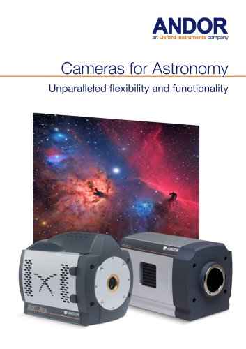
 Cameras for Astronomy
8 Pages
 Microspectroscopy Gatefold
8 Pages
 Spectroscopy Brochure
25 Pages
 iXon EMCCD
13 Pages
 Zyla for Physical Sciences sCMOS
12 Pages
 iVac OEM 2
2 Pages
 iKon-M OEM 2-page PV
2 Pages
 iKon-M X-Ray 2
2 Pages
 Newton EMCCD
16 Pages
 sCMOS
sCMOS16 Pages
 Darkcurrent
3 Pages
 iXon Back-illuminated
6 Pages
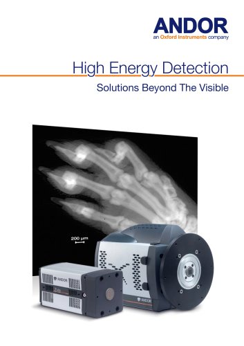 High Energy Detection_2014
28 Pages
 PRODUCT PORTFOLIO 2013
51 Pages
 Neo and Zyla sCMOS
27 Pages
 iXon
iXon33 Pages


Archived catalogs
 Intensified Camera Series
9 Pages
 Clara Interline CCD Series
2 Pages
 Andor Revolution XD brochure
23 Pages
 Revolution DSD
11 Pages
 Clara Flyer
2 Pages
 Revolution
23 Pages
 Low Light Imaging
8 Pages
 iQ Software
12 Pages
 Luca vs Interline CCD
6 Pages
 ApogeeAspen CG9000
5 Pages
 Apogee Aspen CG6
5 Pages
 Apogee Aspen CG47
5 Pages
 Apogee Aspen CG230
5 Pages
 Apogee Alta F9000
5 Pages
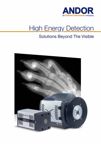 High Energy Detection_2019
8 Pages
 Astronomy Brochure_2014
19 Pages
 Astronomy Brochure
19 Pages
 iKon-M USB X-Ray Brochure
2 Pages


- Bourn And Koch digital camera
- Bourn And Koch visible camera
- Bourn And Koch management software
- Bourn And Koch automation software
- Bourn And Koch CMOS camera
- Bourn And Koch industrial camera
- Bourn And Koch analysis software
- Infrared imager
- Bourn And Koch process software
- Monitoring camera system
- Bourn And Koch full-color camera
- Bourn And Koch Windows software
- Bourn And Koch real-time software
- Bourn And Koch cloud software
- Bourn And Koch control software
- Bourn And Koch spectrometer
- Bourn And Koch USB camera
- Bourn And Koch detection camera
- Bourn And Koch image processing camera